Salloway Stephen, Gamez Jose E, Singh Upinder, Sadowsky Carl H, Villena Teresa, Sabbagh Marwan N, Beach Thomas G, Duara Ranjan, Fleisher Adam S, Frey Kirk A, Walker Zuzana, Hunjan Arvinder, Escovar Yavir M, Agronin Marc E, Ross Joel, Bozoki Andrea, Akinola Mary, Shi Jiong, Vandenberghe Rik, Ikonomovic Milos D, Sherwin Paul F, Farrar Gill, Smith Adrian P L, Buckley Christopher J, Thal Dietmar Rudolf, Zanette Michelle, Curtis Craig
Neurology and the Memory and Aging Program, Butler Hospital, Warren Alpert Medical School, Brown University, Providence, RI, USA.
Department of Neurology and Psychiatry, Warren Alpert Medical School, Brown University, Providence, RI, USA.
Alzheimers Dement (Amst). 2017 Jul 1;9:25-34. doi: 10.1016/j.dadm.2017.06.001. eCollection 2017.
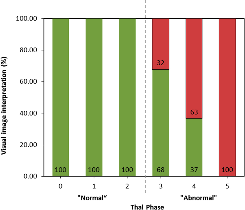
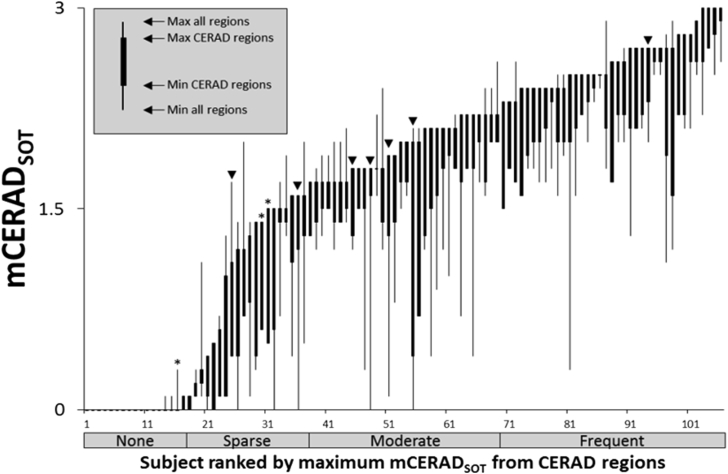
Performance of the amyloid tracer [F]flutemetamol was evaluated against three pathology standard of truth (SoT) measures including neuritic plaques (CERAD "original" and "modified" and the amyloid component of the 2012 NIA-AA guidelines).
After [F]flutemetamol imaging, 106 end-of-life patients who died underwent postmortem brain examination for amyloid plaque load. Blinded positron emission tomography scan interpretations by five independent electronically trained readers were compared with pathology measures.
By SoT, sensitivity and specificity of majority image interpretations were, respectively, 91.9% and 87.5% with "original CERAD," 90.8% and 90.0% with "modified CERAD," and 85.7% and 100% with the 2012 NIA-AA criteria.
The high accuracy of either CERAD criteria suggests that [F]flutemetamol predominantly reflects neuritic amyloid plaque density. However, the use of CERAD criteria as the SoT can result in some false-positive results because of the presence of diffuse plaques, which are accounted for when the positron emission tomography read is compared with the 2012 NIA-AA criteria.
针对三种病理学真值标准(SoT)测量方法,对淀粉样蛋白示踪剂[F]氟代米他莫的性能进行了评估,这三种方法包括神经炎性斑块(CERAD“原始”和“修订”版本以及2012年NIA-AA指南中的淀粉样蛋白成分)。
在进行[F]氟代米他莫成像后,对106例临终死亡患者进行了死后脑淀粉样斑块负荷检查。将五名独立的经过电子培训的阅片者对正电子发射断层扫描的盲法解读结果与病理学测量结果进行比较。
根据SoT标准,大多数图像解读的敏感度和特异度分别为:采用“原始CERAD”时为91.9%和87.5%,采用“修订CERAD”时为90.8%和90.0%,采用2012年NIA-AA标准时为85.7%和100%。
两种CERAD标准的高准确性表明,[F]氟代米他莫主要反映神经炎性淀粉样斑块密度。然而,由于存在弥漫性斑块,将CERAD标准用作SoT可能会导致一些假阳性结果,而在将正电子发射断层扫描解读结果与2012年NIA-AA标准进行比较时会考虑到这些弥漫性斑块。