Steinzeig Anna, Molotkov Dmitry, Castrén Eero
Neuroscience center, University of Helsinki, Helsinki, Finland.
Biomedicum Imaging Unit, University of Helsinki, Helsinki, Finland.
PLoS One. 2017 Aug 16;12(8):e0181788. doi: 10.1371/journal.pone.0181788. eCollection 2017.
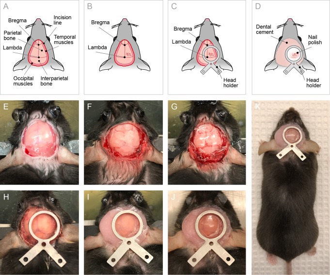
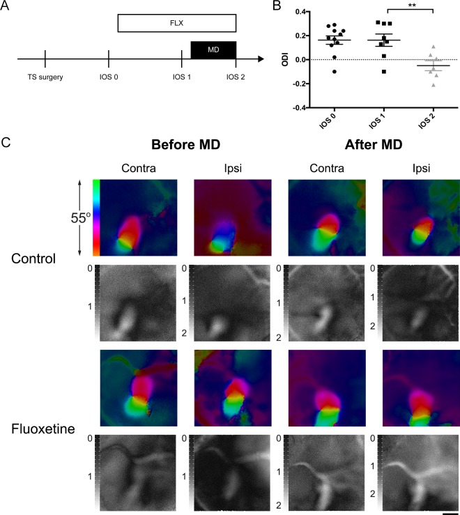
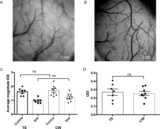
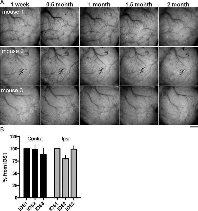
Growing interest in long-term visualization of cortical structure and function requires methods that allow observation of an intact cortex in longitudinal imaging studies. Here we describe a detailed protocol for the "transparent skull" (TS) preparation based on skull clearing with cyanoacrylate, which is applicable for long-term imaging through the intact skull in mice. We characterized the properties of the TS in imaging of intrinsic optical signals and compared them with the more conventional cranial window preparation. Our results show that TS is less invasive, maintains stabile transparency for at least two months, and compares favorably to data obtained from the conventional cranial window. We applied this method to experiments showing that a four-week treatment with the antidepressant fluoxetine combined with one week of monocular deprivation induced a shift in ocular dominance in the mouse visual cortex, confirming that fluoxetine treatment restores critical-period-like plasticity. Our results demonstrate that the TS preparation could become a useful method for long-term visualization of the living mouse brain.
对皮质结构和功能进行长期可视化的兴趣日益浓厚,这就需要在纵向成像研究中采用能够观察完整皮质的方法。在此,我们描述了一种基于用氰基丙烯酸酯进行颅骨清理的“透明颅骨”(TS)制备的详细方案,该方案适用于通过完整颅骨对小鼠进行长期成像。我们在固有光信号成像中表征了TS的特性,并将其与更传统的颅骨窗制备方法进行了比较。我们的结果表明,TS的侵入性较小,至少能保持两个月的稳定透明度,并且与从传统颅骨窗获得的数据相比具有优势。我们将此方法应用于实验,结果显示用抗抑郁药氟西汀进行四周治疗并结合一周的单眼剥夺会导致小鼠视觉皮质中眼优势的转变,证实氟西汀治疗可恢复类似关键期的可塑性。我们的结果表明,TS制备可能成为长期可视化活体小鼠大脑的一种有用方法。