Hong Jinsung, Murugesan Sricharan, Betzig Eric, Hammer John A
Cell Biology and Physiology Center, National Heart, Lung and Blood Institute, National Institutes of Health, Bethesda, MD, United States of America.
Janelia Research Campus/HHMI, Ashburn, VA, United States of America.
PLoS One. 2017 Aug 17;12(8):e0183174. doi: 10.1371/journal.pone.0183174. eCollection 2017.
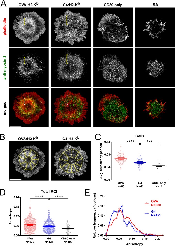
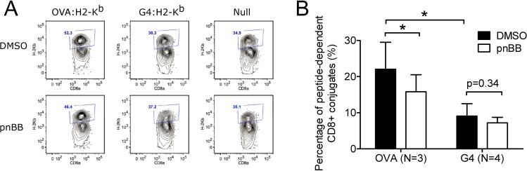
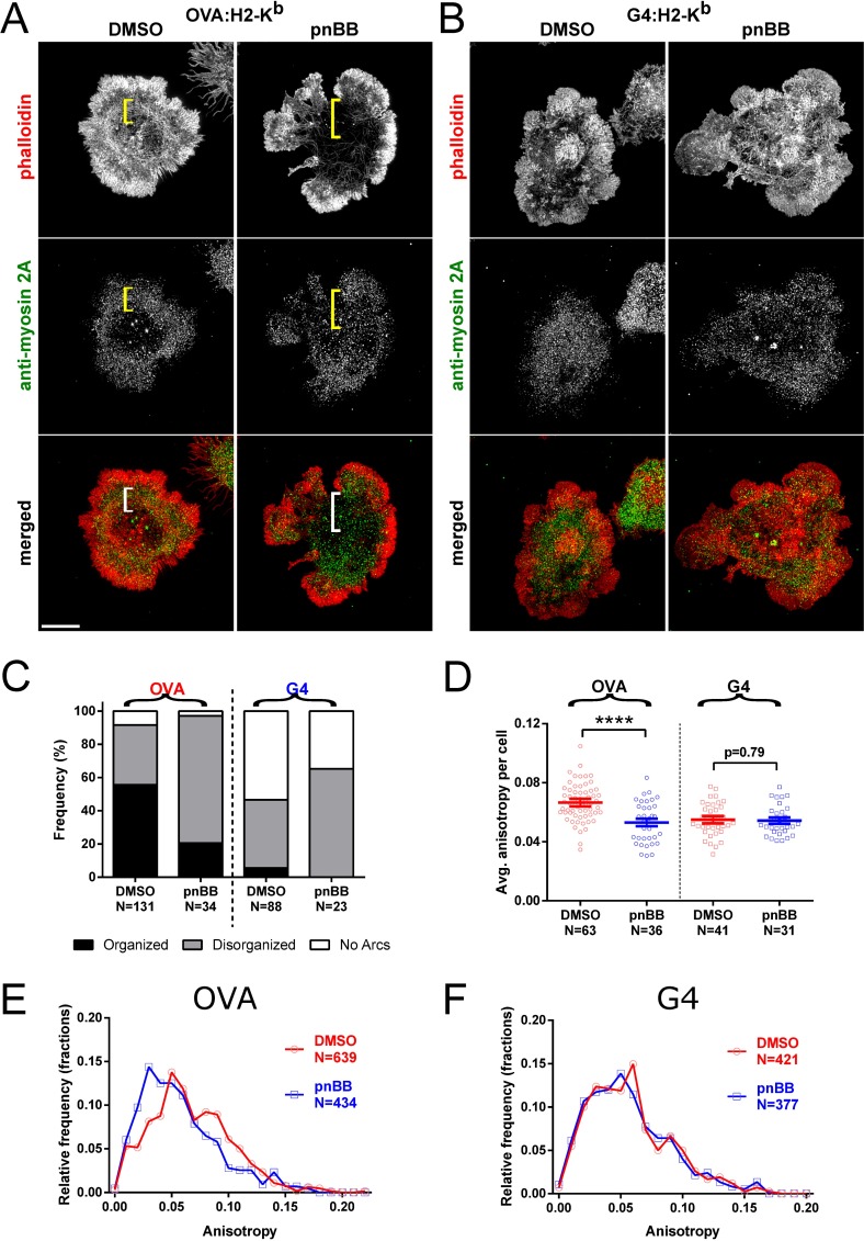
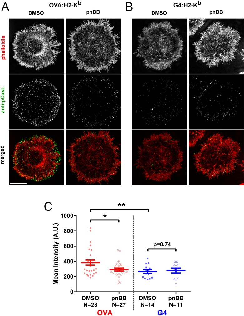
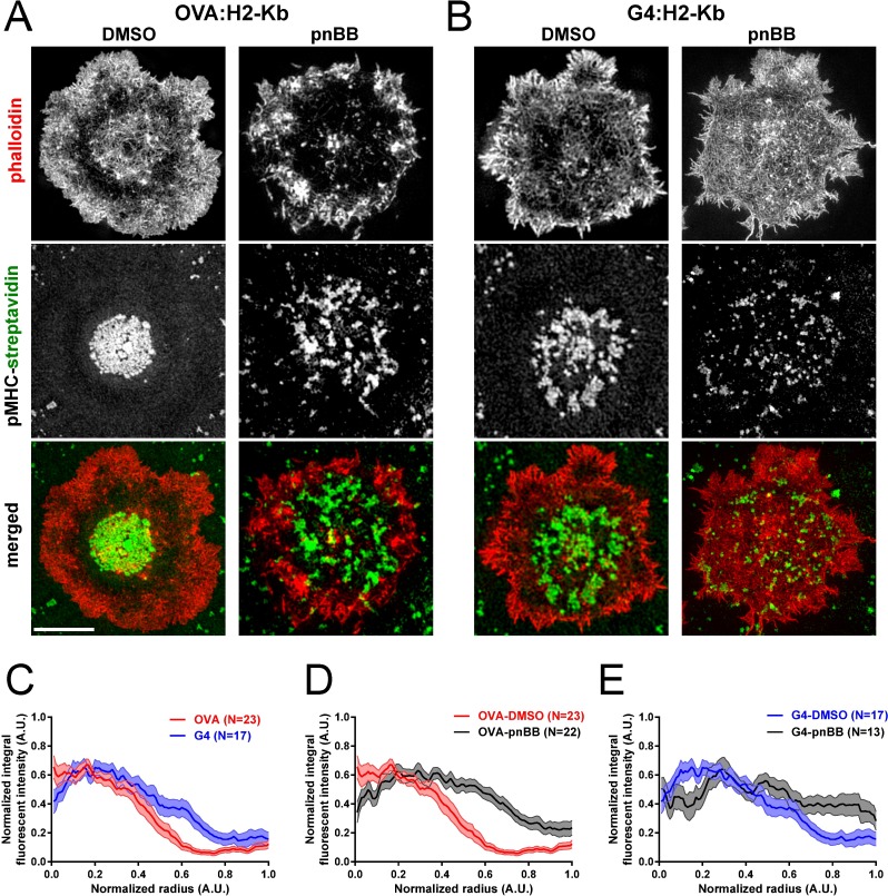
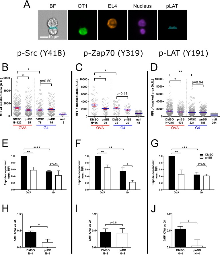
Mechano-transduction is an emerging but still poorly understood component of T cell activation. Here we investigated the ligand-dependent contribution made by contractile actomyosin arcs populating the peripheral supramolecular activation cluster (pSMAC) region of the immunological synapse (IS) to T cell receptor (TCR) microcluster transport and proximal signaling in primary mouse T cells. Using super resolution microscopy, OT1-CD8+ mouse T cells, and two ovalbumin (OVA) peptides with different affinities for the TCR, we show that the generation of organized actomyosin arcs depends on ligand potency and the ability of myosin 2 to contract actin filaments. While weak ligands induce disorganized actomyosin arcs, strong ligands result in organized actomyosin arcs that correlate well with tension-sensitive CasL phosphorylation and the accumulation of ligands at the IS center. Blocking myosin 2 contractility greatly reduces the difference in the extent of Src and LAT phosphorylation observed between the strong and the weak ligand, arguing that myosin 2-dependent force generation within actin arcs contributes to ligand discrimination. Together, our data are consistent with the idea that actomyosin arcs in the pSMAC region of the IS promote a mechano-chemical feedback mechanism that amplifies the accumulation of critical signaling molecules at the IS.
机械转导是T细胞活化中一个新兴但仍未被充分理解的组成部分。在这里,我们研究了收缩性肌动球蛋白弧对原发性小鼠T细胞中T细胞受体(TCR)微簇转运和近端信号传导的配体依赖性贡献,这些收缩性肌动球蛋白弧分布在免疫突触(IS)的外周超分子活化簇(pSMAC)区域。使用超分辨率显微镜、OT1-CD8 +小鼠T细胞以及两种对TCR具有不同亲和力的卵清蛋白(OVA)肽,我们发现有组织的肌动球蛋白弧的产生取决于配体效力和肌球蛋白2收缩肌动蛋白丝的能力。弱配体诱导无序的肌动球蛋白弧,而强配体则导致有组织的肌动球蛋白弧,这与张力敏感的CasL磷酸化以及配体在IS中心的积累密切相关。阻断肌球蛋白2的收缩性会大大降低强配体和弱配体之间Src和LAT磷酸化程度的差异,这表明肌动蛋白弧内肌球蛋白2依赖性力的产生有助于配体识别。总之,我们的数据与以下观点一致,即IS的pSMAC区域中的肌动球蛋白弧促进了一种机械化学反馈机制,该机制放大了关键信号分子在IS处的积累。