Ariza Jeanelle, Hurtado Jesus, Rogers Haille, Ikeda Raymond, Dill Michael, Steward Craig, Creary Donnay, Van de Water Judy, Martínez-Cerdeño Verónica
Department of Pathology and Laboratory Medicine, Sacramento, CA, United States of America.
Institute for Pediatric Regenerative Medicine and Shriners Hospitals for Children Northern California, Sacramento, CA, United States of America.
PLoS One. 2017 Aug 18;12(8):e0183443. doi: 10.1371/journal.pone.0183443. eCollection 2017.
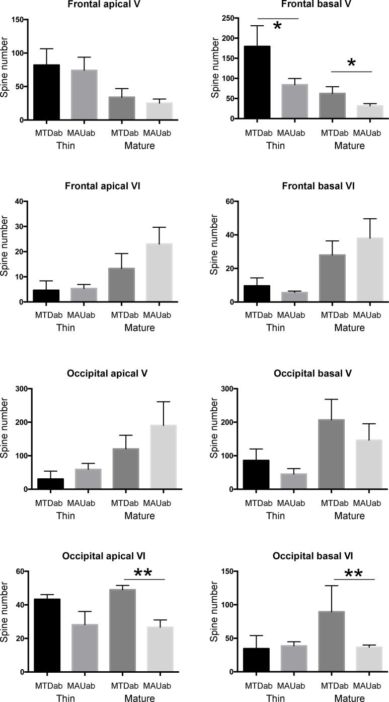
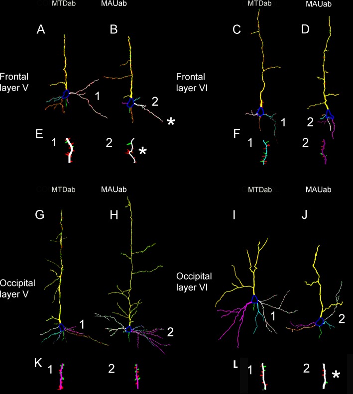
An association between maternal IgG antibodies reactive against proteins in fetal brain and an outcome of autism in the child has been identified. Using a mouse model of prenatal intraventricular administration of autism-specific maternal IgG, we demonstrated that these antibodies produce behavioral alterations similar to those in children with ASD. We previously demonstrated that these antibodies bind to radial glial stem cells (RG) and observed an increase in the number of divisions of translocating RG in the developing cortex. We also showed an alteration in brain size and as well as a generalized increased of neuronal volume in adult mice. Here, we used our intraventricular mouse model of antibody administration, followed by Golgi and Neurolucida analysis to demonstrate that during midstages of neurogenesis these maternal autism-specific antibodies produced a consistent decrease in the number of spines in the infragranular layers in the adult cortical areas analyzed. Specifically, in the frontal cortex basal dendrites of layer V neurons were decreased in length and volume, and both the total number of spines-mature and immature-and the spine density were lower than in the control neurons from the same region. Further, in the occipital cortex layer VI neurons presented with a decrease in the total number of spines and in the spine density in the apical dendrite, as well as decrease in the number of mature spines in the apical and basal dendrites. Interestingly, the time of exposure to these antibodies (E14.5) coincides with the generation of pyramidal neurons in layer V in the frontal cortex and in layer VI in the occipital cortex, following the normal rostro-caudal pattern of cortical cell generation. We recently demonstrated that one of the primary antigens recognized by these antibodies corresponds to stress-induced phosphoprotein 1 (STIP1). Here we hypothesize that the reduction in the access of newborn cells to STIP1 in the developing cortex may be responsible for the reduced dendritic arborization and number of spines we noted in the adult cortex.
已确定母体针对胎儿脑内蛋白质的IgG抗体与儿童自闭症结局之间存在关联。利用产前脑室内注射自闭症特异性母体IgG的小鼠模型,我们证明这些抗体产生的行为改变与自闭症谱系障碍(ASD)儿童相似。我们之前证明这些抗体与放射状胶质干细胞(RG)结合,并观察到发育中的皮质中迁移RG的分裂数量增加。我们还显示成年小鼠脑大小改变以及神经元体积普遍增大。在此,我们利用脑室内抗体注射小鼠模型,随后进行高尔基染色和Neurolucida分析,以证明在神经发生中期,这些母体自闭症特异性抗体在分析的成年皮质区域颗粒下层中产生棘突数量持续减少。具体而言,在额叶皮质,V层神经元的基底树突长度和体积减小,成熟和未成熟棘突的总数以及棘突密度均低于同一区域的对照神经元。此外,在枕叶皮质,VI层神经元的顶树突棘突总数和棘突密度降低,顶树突和基底树突中的成熟棘突数量也减少。有趣的是,暴露于这些抗体的时间(E14.5)与额叶皮质V层和枕叶皮质VI层锥体神经元的生成时间一致,并遵循皮质细胞生成的正常头-尾模式。我们最近证明这些抗体识别的主要抗原之一对应于应激诱导磷蛋白1(STIP1)。在此我们假设,发育中的皮质中新生细胞接触STIP1减少可能是我们在成年皮质中观察到的树突分支减少和棘突数量减少的原因。