• 文献检索
  • 文档翻译
  • 深度研究
  • 学术资讯
  • Suppr Zotero 插件Zotero 插件
  • 邀请有礼
  • 套餐&价格
  • 历史记录
应用&插件
Suppr Zotero 插件Zotero 插件浏览器插件Mac 客户端Windows 客户端微信小程序
定价
高级版会员购买积分包购买API积分包
服务
文献检索文档翻译深度研究API 文档MCP 服务
关于我们
关于 Suppr公司介绍联系我们用户协议隐私条款
关注我们

Suppr 超能文献

核心技术专利:CN118964589B侵权必究
粤ICP备2023148730 号-1Suppr @ 2026

文献检索

告别复杂PubMed语法,用中文像聊天一样搜索,搜遍4000万医学文献。AI智能推荐,让科研检索更轻松。

立即免费搜索

文件翻译

保留排版,准确专业,支持PDF/Word/PPT等文件格式,支持 12+语言互译。

免费翻译文档

深度研究

AI帮你快速写综述,25分钟生成高质量综述,智能提取关键信息,辅助科研写作。

立即免费体验

一种由顶端MRCK驱动的形态发生途径控制上皮极性。

An apical MRCK-driven morphogenetic pathway controls epithelial polarity.

作者信息

Zihni Ceniz, Vlassaks Evi, Terry Stephen, Carlton Jeremy, Leung Thomas King Chor, Olson Michael, Pichaud Franck, Balda Maria Susana, Matter Karl

机构信息

Institute of Ophthalmology, University College London, Bath Street, London EC1V 9EL, UK.

MRC Laboratory for Molecular Cell Biology, University College London, Gower Street, London WC1E 6BT, UK.

出版信息

Nat Cell Biol. 2017 Sep;19(9):1049-1060. doi: 10.1038/ncb3592. Epub 2017 Aug 21.

DOI:10.1038/ncb3592
PMID:28825699
原文链接:https://pmc.ncbi.nlm.nih.gov/articles/PMC5617103/
Abstract

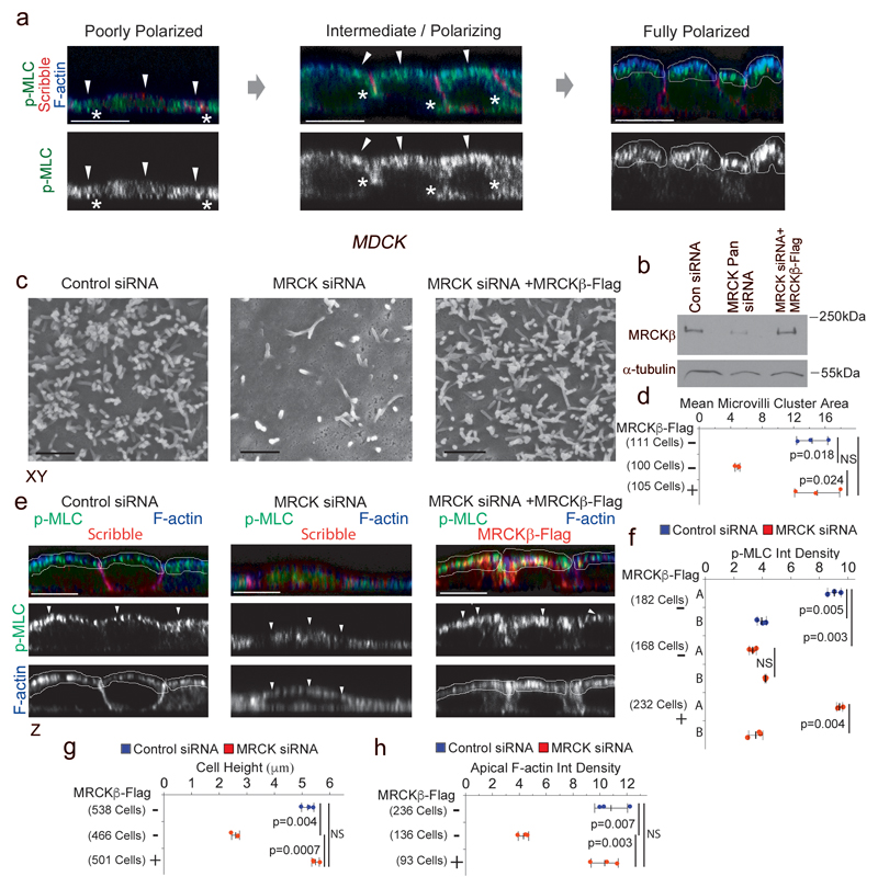
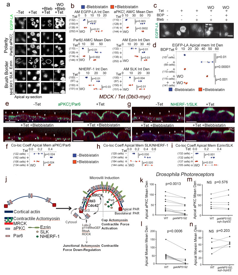
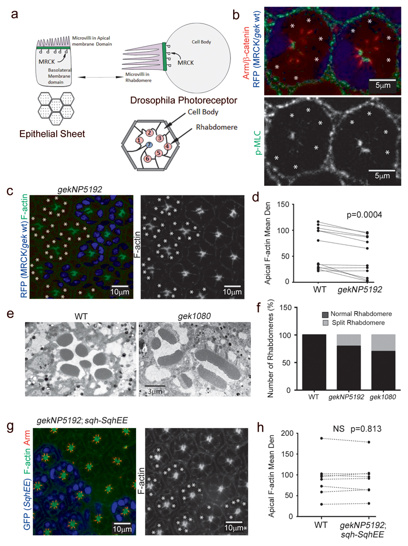
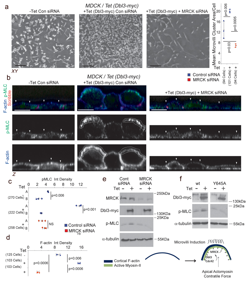
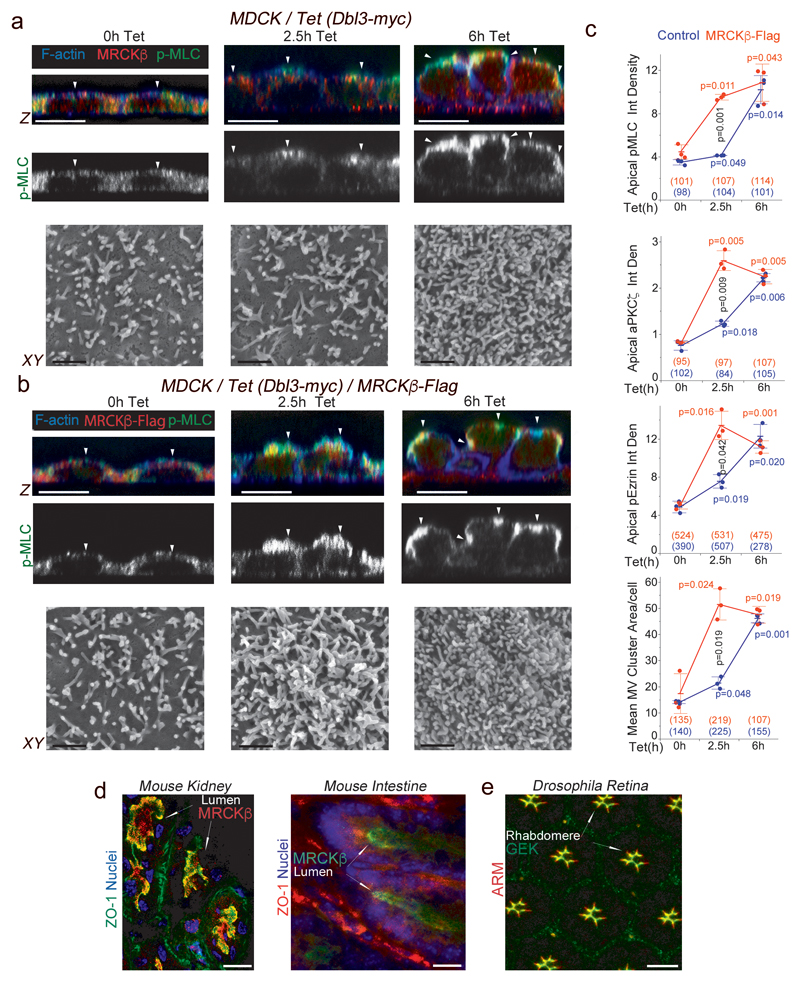
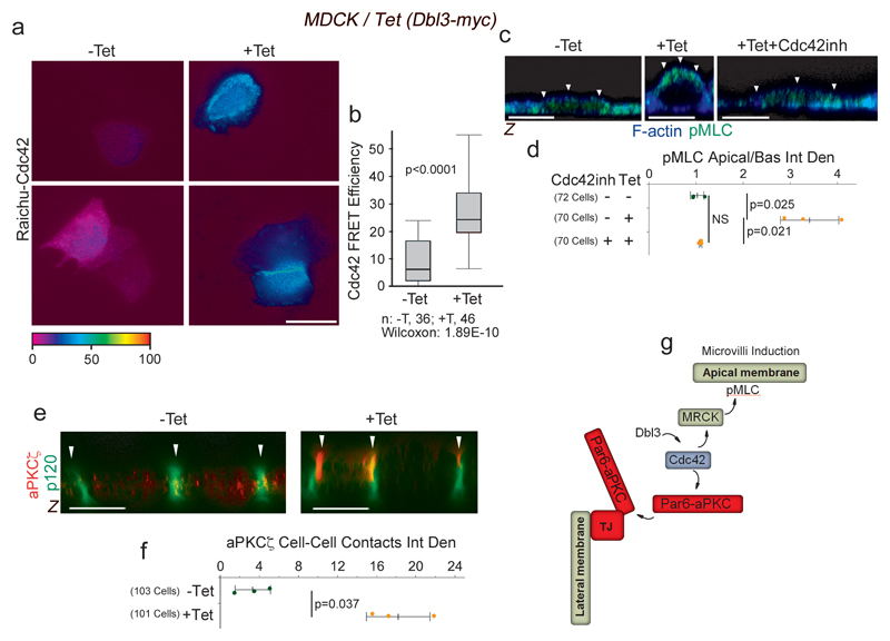
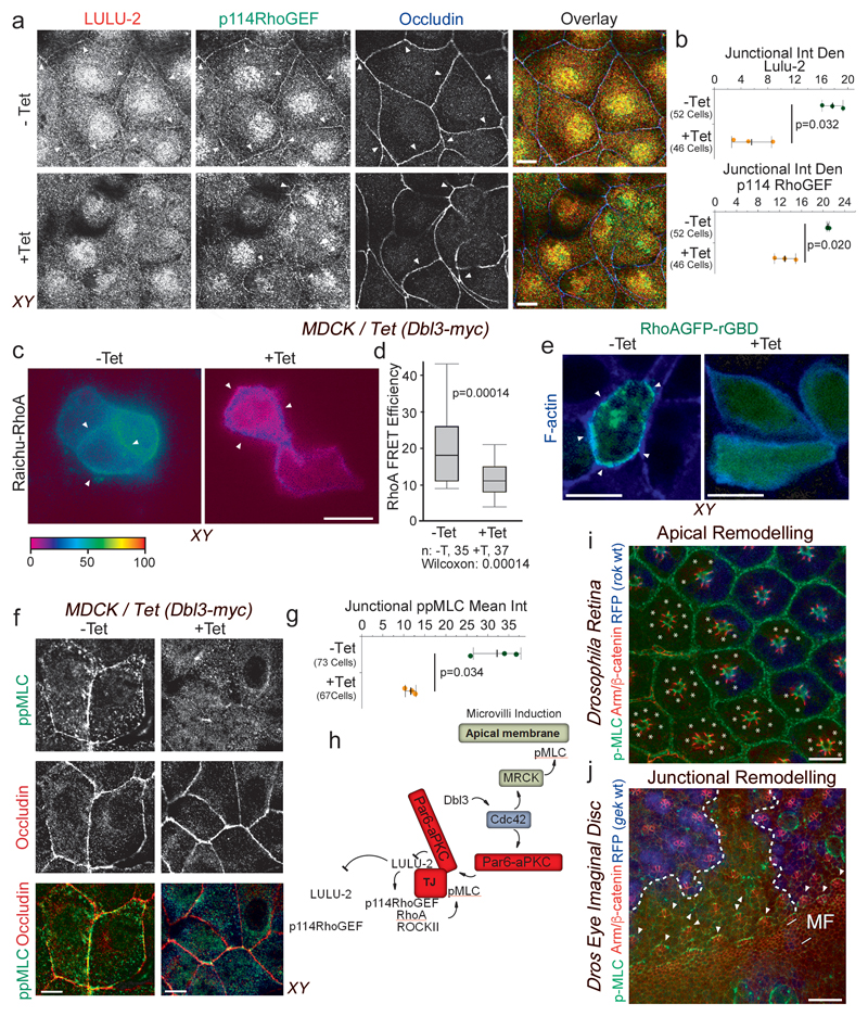
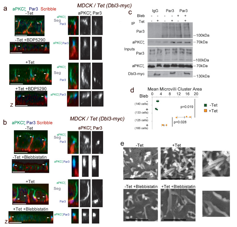
Polarized epithelia develop distinct cell surface domains, with the apical membrane acquiring characteristic morphological features such as microvilli. Cell polarization is driven by polarity determinants including the evolutionarily conserved partitioning-defective (PAR) proteins that are separated into distinct cortical domains. PAR protein segregation is thought to be a consequence of asymmetric actomyosin contractions. The mechanism of activation of apically polarized actomyosin contractility is unknown. Here we show that the Cdc42 effector MRCK activates myosin-II at the apical pole to segregate aPKC-Par6 from junctional Par3, defining the apical domain. Apically polarized MRCK-activated actomyosin contractility is reinforced by cooperation with aPKC-Par6 downregulating antagonistic RhoA-driven junctional actomyosin contractility, and drives polarization of cytosolic brush border determinants and apical morphogenesis. MRCK-activated polarized actomyosin contractility is required for apical differentiation and morphogenesis in vertebrate epithelia and Drosophila photoreceptors. Our results identify an apical origin of actomyosin-driven morphogenesis that couples cytoskeletal reorganization to PAR polarity signalling.

摘要

极化上皮细胞形成不同的细胞表面结构域,顶端膜具有微绒毛等特征性形态特征。细胞极化由极性决定因子驱动,包括进化上保守的分区缺陷(PAR)蛋白,这些蛋白被分隔到不同的皮质结构域。PAR蛋白的分离被认为是肌动球蛋白不对称收缩的结果。顶端极化的肌动球蛋白收缩性的激活机制尚不清楚。在这里,我们表明Cdc42效应器MRCK在顶端极激活肌球蛋白-II,将aPKC-Par6与连接蛋白Par3分离,从而定义顶端结构域。顶端极化的MRCK激活的肌动球蛋白收缩性通过与aPKC-Par6协同作用而增强,aPKC-Par6下调拮抗的RhoA驱动的连接肌动球蛋白收缩性,并驱动细胞质刷状缘决定因子的极化和顶端形态发生。MRCK激活的极化肌动球蛋白收缩性是脊椎动物上皮细胞和果蝇光感受器顶端分化和形态发生所必需的。我们的结果确定了肌动球蛋白驱动的形态发生的顶端起源,它将细胞骨架重组与PAR极性信号联系起来。

https://cdn.ncbi.nlm.nih.gov/pmc/blobs/416a/5617103/2bdb00a8e161/emss-73446-f008.jpg
https://cdn.ncbi.nlm.nih.gov/pmc/blobs/416a/5617103/aa2f6a12cbe7/emss-73446-f001.jpg
https://cdn.ncbi.nlm.nih.gov/pmc/blobs/416a/5617103/03f6b67833bf/emss-73446-f002.jpg
https://cdn.ncbi.nlm.nih.gov/pmc/blobs/416a/5617103/0fe96aa239c4/emss-73446-f003.jpg
https://cdn.ncbi.nlm.nih.gov/pmc/blobs/416a/5617103/93ce08c4a6e8/emss-73446-f004.jpg
https://cdn.ncbi.nlm.nih.gov/pmc/blobs/416a/5617103/e8394e4c29ec/emss-73446-f005.jpg
https://cdn.ncbi.nlm.nih.gov/pmc/blobs/416a/5617103/7582cb6e31e0/emss-73446-f006.jpg
https://cdn.ncbi.nlm.nih.gov/pmc/blobs/416a/5617103/47a7726c958c/emss-73446-f007.jpg
https://cdn.ncbi.nlm.nih.gov/pmc/blobs/416a/5617103/2bdb00a8e161/emss-73446-f008.jpg

相似文献

1
An apical MRCK-driven morphogenetic pathway controls epithelial polarity.一种由顶端MRCK驱动的形态发生途径控制上皮极性。
Nat Cell Biol. 2017 Sep;19(9):1049-1060. doi: 10.1038/ncb3592. Epub 2017 Aug 21.
2
The WD40 protein Morg1 facilitates Par6-aPKC binding to Crb3 for apical identity in epithelial cells.WD40 蛋白 Morg1 促进 Par6-aPKC 与 Crb3 的结合,以维持上皮细胞的顶端身份。
J Cell Biol. 2013 Mar 4;200(5):635-50. doi: 10.1083/jcb.201208150. Epub 2013 Feb 25.
3
Dbl3 drives Cdc42 signaling at the apical margin to regulate junction position and apical differentiation.Dbl3 在顶端边缘驱动 Cdc42 信号传导,以调节连接位置和顶端分化。
J Cell Biol. 2014 Jan 6;204(1):111-27. doi: 10.1083/jcb.201304064. Epub 2013 Dec 30.
4
Cdc42 defines apical identity and regulates epithelial morphogenesis by promoting apical recruitment of Par6-aPKC and Crumbs.Cdc42 通过促进 Par6-aPKC 和 Crumbs 在上皮细胞的顶端募集来定义顶端身份并调节上皮形态发生。
Development. 2019 Aug 12;146(15):dev175497. doi: 10.1242/dev.175497.
5
Pak1 Kinase Maintains Apical Membrane Identity in Epithelia.Pak1 激酶维持上皮细胞的顶端膜特性。
Cell Rep. 2018 Feb 13;22(7):1639-1646. doi: 10.1016/j.celrep.2018.01.060.
6
A polybasic domain in aPKC mediates Par6-dependent control of membrane targeting and kinase activity.多碱性结构域在 aPKC 中介导 Par6 依赖性的膜靶向和激酶活性的调控。
J Cell Biol. 2020 Jul 6;219(7). doi: 10.1083/jcb.201903031.
7
The Cdc42/Par6/aPKC polarity complex regulates apoptosis-induced compensatory proliferation in epithelia.Cdc42/Par6/aPKC 极性复合物调节上皮细胞凋亡诱导的代偿性增殖。
Curr Biol. 2010 Apr 27;20(8):677-86. doi: 10.1016/j.cub.2010.03.025. Epub 2010 Apr 8.
8
Par-3 family proteins in cell polarity & adhesion.细胞极性和黏附中的Par-3家族蛋白。
FEBS J. 2022 Feb;289(3):596-613. doi: 10.1111/febs.15754. Epub 2021 Mar 3.
9
Willin and Par3 cooperatively regulate epithelial apical constriction through aPKC-mediated ROCK phosphorylation.威尔蛋白和 Par3 通过蛋白激酶 C 介导的 ROCK 磷酸化协同调节上皮细胞顶端缢缩。
Nat Cell Biol. 2011 Jun 19;13(7):860-6. doi: 10.1038/ncb2274.
10
DAPLE orchestrates apical actomyosin assembly from junctional polarity complexes.DAPLE 从连接极性复合物中协调顶端肌动球蛋白的组装。
J Cell Biol. 2022 May 2;221(5). doi: 10.1083/jcb.202111002. Epub 2022 Apr 7.

引用本文的文献

1
Phagocytosis by the retinal pigment epithelium: New insights into polarized cell mechanics.视网膜色素上皮细胞的吞噬作用:极化细胞力学的新见解
Bioessays. 2025 Jan;47(1):e2300197. doi: 10.1002/bies.202300197. Epub 2024 Dec 11.
2
Loss of Cdc42 causes abnormal optic cup morphogenesis and microphthalmia in mouse.Cdc42缺失导致小鼠视杯形态发生异常和小眼畸形。
Front Cell Neurosci. 2024 Nov 20;18:1474010. doi: 10.3389/fncel.2024.1474010. eCollection 2024.
3
MRCK-1 activates non-muscle myosin for outgrowth of a unicellular tube in Caenorhabditis elegans.

本文引用的文献

1
Tight junctions: from simple barriers to multifunctional molecular gates.紧密连接:从简单的屏障到多功能分子门控。
Nat Rev Mol Cell Biol. 2016 Sep;17(9):564-80. doi: 10.1038/nrm.2016.80. Epub 2016 Jun 29.
2
Pak4 Is Required during Epithelial Polarity Remodeling through Regulating AJ Stability and Bazooka Retention at the ZA.Pak4在通过调节黏附连接稳定性和将Bazooka保留在黏着连接处来重塑上皮极性的过程中是必需的。
Cell Rep. 2016 Apr 5;15(1):45-53. doi: 10.1016/j.celrep.2016.03.014. Epub 2016 Mar 24.
3
Structure, regulation, and functional diversity of microvilli on the apical domain of epithelial cells.
MRCK-1 激活非肌肉肌球蛋白以促进秀丽隐杆线虫单细胞管的延伸。
Development. 2024 Dec 1;151(23). doi: 10.1242/dev.202772. Epub 2024 Nov 29.
4
Loss of Cdc42 causes abnormal optic cup morphogenesis and microphthalmia in mouse.Cdc42缺失导致小鼠视杯形态发生异常和小眼畸形。
bioRxiv. 2024 Oct 22:2024.10.20.619331. doi: 10.1101/2024.10.20.619331.
5
RhoGDI1 regulates cell-cell junctions in polarized epithelial cells.RhoGDI1调节极化上皮细胞中的细胞间连接。
Front Cell Dev Biol. 2024 Jul 17;12:1279723. doi: 10.3389/fcell.2024.1279723. eCollection 2024.
6
RhoA GEF Mcf2lb regulates rosette integrity during collective cell migration.RhoA GEF Mcf2lb 在细胞集体迁移过程中调节玫瑰花结的完整性。
Development. 2024 Jan 1;151(1). doi: 10.1242/dev.201898. Epub 2024 Jan 2.
7
PTPN13 Participates in the Regulation of Epithelial-Mesenchymal Transition and Platinum Sensitivity in High-Grade Serous Ovarian Carcinoma Cells.PTPN13 参与调控高级别浆液性卵巢癌细胞的上皮-间质转化和铂类敏感性。
Int J Mol Sci. 2023 Oct 21;24(20):15413. doi: 10.3390/ijms242015413.
8
MRCK activates mouse oocyte myosin II for spindle rotation and male pronucleus centration.MRCK 激活小鼠卵母细胞肌球蛋白 II 以实现纺锤体旋转和雄性原核中心定位。
J Cell Biol. 2023 Nov 6;222(11). doi: 10.1083/jcb.202211029. Epub 2023 Aug 31.
9
Branched-chain actin dynamics polarizes vesicle trajectories and partitions apicobasal epithelial membrane domains.支链肌动蛋白动力学使囊泡轨迹极化,并分隔顶端基底上皮膜域。
Sci Adv. 2023 Jun 28;9(26):eade4022. doi: 10.1126/sciadv.ade4022.
10
Opportunities and Challenges for the Development of MRCK Kinases Inhibitors as Potential Cancer Chemotherapeutics.MRCK 激酶抑制剂作为潜在癌症化疗药物的发展机遇与挑战。
Cells. 2023 Feb 7;12(4):534. doi: 10.3390/cells12040534.
上皮细胞顶域微绒毛的结构、调控和功能多样性。
Annu Rev Cell Dev Biol. 2015;31:593-621. doi: 10.1146/annurev-cellbio-100814-125234.
4
Shaping the intestinal brush border.塑造肠道刷状缘。
J Cell Biol. 2014 Nov 24;207(4):441-51. doi: 10.1083/jcb.201407015.
5
A novel small-molecule MRCK inhibitor blocks cancer cell invasion.一种新型小分子 MRCK 抑制剂可阻断癌细胞侵袭。
Cell Commun Signal. 2014 Oct 5;12:54. doi: 10.1186/s12964-014-0054-x.
6
Signalling at tight junctions during epithelial differentiation and microbial pathogenesis.上皮细胞分化和微生物致病过程中紧密连接的信号传导
J Cell Sci. 2014 Aug 15;127(Pt 16):3401-13. doi: 10.1242/jcs.145029.
7
The actin-myosin regulatory MRCK kinases: regulation, biological functions and associations with human cancer.肌动球蛋白调节性 MRCK 激酶:调节、生物学功能及与人类癌症的关联。
J Mol Med (Berl). 2014 Mar;92(3):217-25. doi: 10.1007/s00109-014-1133-6. Epub 2014 Feb 20.
8
Dynamics of ezrin and EBP50 in regulating microvilli on the apical aspect of epithelial cells.上皮细胞顶侧微绒毛的 ezrin 和 EBP50 调节动力学。
Biochem Soc Trans. 2014 Feb;42(1):189-94. doi: 10.1042/BST20130263.
9
Dbl3 drives Cdc42 signaling at the apical margin to regulate junction position and apical differentiation.Dbl3 在顶端边缘驱动 Cdc42 信号传导,以调节连接位置和顶端分化。
J Cell Biol. 2014 Jan 6;204(1):111-27. doi: 10.1083/jcb.201304064. Epub 2013 Dec 30.
10
Principles of PAR polarity in Caenorhabditis elegans embryos.线虫胚胎中 PAR 极性的原理。
Nat Rev Mol Cell Biol. 2013 May;14(5):315-22. doi: 10.1038/nrm3558. Epub 2013 Apr 18.