• 文献检索
  • 文档翻译
  • 深度研究
  • 学术资讯
  • Suppr Zotero 插件Zotero 插件
  • 邀请有礼
  • 套餐&价格
  • 历史记录
应用&插件
Suppr Zotero 插件Zotero 插件浏览器插件Mac 客户端Windows 客户端微信小程序
定价
高级版会员购买积分包购买API积分包
服务
文献检索文档翻译深度研究API 文档MCP 服务
关于我们
关于 Suppr公司介绍联系我们用户协议隐私条款
关注我们

Suppr 超能文献

核心技术专利:CN118964589B侵权必究
粤ICP备2023148730 号-1Suppr @ 2026

文献检索

告别复杂PubMed语法,用中文像聊天一样搜索,搜遍4000万医学文献。AI智能推荐,让科研检索更轻松。

立即免费搜索

文件翻译

保留排版,准确专业,支持PDF/Word/PPT等文件格式,支持 12+语言互译。

免费翻译文档

深度研究

AI帮你快速写综述,25分钟生成高质量综述,智能提取关键信息,辅助科研写作。

立即免费体验

掌腱膜挛缩症中类似胚胎干细胞的细胞群表达肾素-血管紧张素系统的成分。

Embryonic Stem Cell-Like Population in Dupuytren's Disease Expresses Components of the Renin-Angiotensin System.

作者信息

On Nicholas, Koh Sabrina P, Brasch Helen D, Dunne Jonathan C, Armstrong James R, Tan Swee T, Itinteang Tinte

机构信息

Gillies McIndoe Research Institute; and Wellington Regional Plastic, Reconstructive, Maxillofacial and Burns Unit, Hutt Hospital, Wellington, New Zealand.

出版信息

Plast Reconstr Surg Glob Open. 2017 Jul 24;5(7):e1422. doi: 10.1097/GOX.0000000000001422. eCollection 2017 Jul.

DOI:10.1097/GOX.0000000000001422
PMID:28831359
原文链接:https://pmc.ncbi.nlm.nih.gov/articles/PMC5548582/
Abstract

UNLABELLED

The renin-angiotensin system (RAS) mediates cardiac and renal fibrosis. Dupuytren's disease (DD) is a proliferative fibromatosis affecting the hands. This study investigated the expression of the RAS in DD.

METHODS

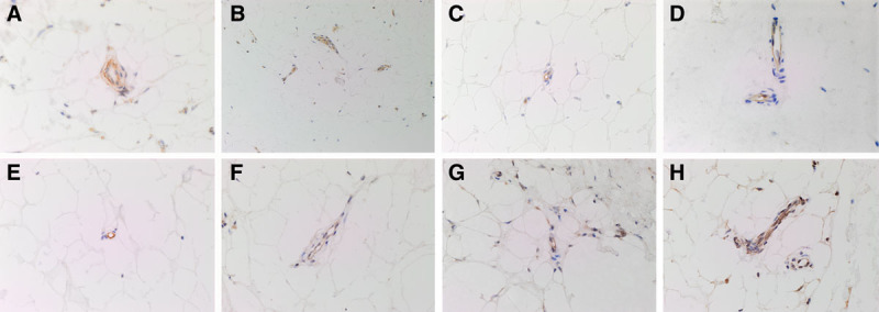
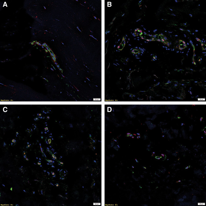
3,3-Diaminobenzidine (DAB) and immunofluorescent immunohistochemical (IHC) staining for (pro)renin receptor (PRR), angiotensin-converting enzyme (ACE), angiotensin II receptor 1 (ATIIR1), and angiotensin II receptor 2 (ATIIR2) was performed on 4-μm thick formalin-fixed paraffin-embedded sections of DD cords and nodules from 6 patients. Western blotting (WB) and NanoString mRNA analysis were performed to confirm RAS protein expression and transcriptional activation, respectively.

RESULTS

IHC staining demonstrated the expression of PRR, ACE, ATIIR1, and ATIIR2 on the ERG and CD34 endothelium of the micro vessels surrounding the DD cords and nodules. PRR was also expressed on the pericyte layer of these microvessels. WB confirmed protein expression of PRR, ACE, and ATIIR2 but not ATIIR1. NanoString analysis confirmed transcriptional activation of PRR, ACE, ATIIR1, but ATIIR2 was below detectable levels.

CONCLUSIONS

We demonstrated expression of PRR, ATIIR1, ATIIR2, and ACE on the embryonic stem cell-like cell population on the microvessels surrounding DD nodules and cords by IHC staining, although the expression of ATIIR1 was not confirmed by WB and that of ATIIR2 was below detectable levels on NanoString analysis. These findings suggest the embryonic stem cell-like cell population as a potential therapeutic target for DD, by using RAS modulators.

摘要

未标记

肾素-血管紧张素系统(RAS)介导心脏和肾脏纤维化。杜普伊特伦挛缩病(DD)是一种影响手部的增生性纤维瘤病。本研究调查了RAS在DD中的表达。

方法

对6例患者的DD条索和结节的4μm厚福尔马林固定石蜡包埋切片进行3,3-二氨基联苯胺(DAB)和免疫荧光免疫组织化学(IHC)染色,检测(前)肾素受体(PRR)、血管紧张素转换酶(ACE)、血管紧张素II受体1(ATIIR1)和血管紧张素II受体2(ATIIR2)。分别进行蛋白质印迹法(WB)和NanoString mRNA分析以确认RAS蛋白表达和转录激活。

结果

免疫组织化学染色显示PRR、ACE、ATIIR1和ATIIR2在DD条索和结节周围微血管的ERG和CD34内皮细胞上表达。PRR也在这些微血管的周细胞层表达。蛋白质印迹法证实了PRR、ACE和ATIIR2的蛋白表达,但未证实ATIIR1的表达。NanoString分析证实了PRR、ACE、ATIIR1的转录激活,但ATIIR2低于可检测水平。

结论

我们通过免疫组织化学染色证明了PRR、ATIIR1、ATIIR2和ACE在DD结节和条索周围微血管上的胚胎干细胞样细胞群体中的表达,尽管蛋白质印迹法未证实ATIIR1的表达,且NanoString分析显示ATIIR2低于可检测水平。这些发现表明,通过使用RAS调节剂,胚胎干细胞样细胞群体可能是DD的潜在治疗靶点。

https://cdn.ncbi.nlm.nih.gov/pmc/blobs/16c5/5548582/89044738a8f0/gox-5-e1422-g004.jpg
https://cdn.ncbi.nlm.nih.gov/pmc/blobs/16c5/5548582/914d828ca897/gox-5-e1422-g001.jpg
https://cdn.ncbi.nlm.nih.gov/pmc/blobs/16c5/5548582/c65d7c89c977/gox-5-e1422-g002.jpg
https://cdn.ncbi.nlm.nih.gov/pmc/blobs/16c5/5548582/ea566f5dd72b/gox-5-e1422-g003.jpg
https://cdn.ncbi.nlm.nih.gov/pmc/blobs/16c5/5548582/89044738a8f0/gox-5-e1422-g004.jpg

相似文献

1
Embryonic Stem Cell-Like Population in Dupuytren's Disease Expresses Components of the Renin-Angiotensin System.掌腱膜挛缩症中类似胚胎干细胞的细胞群表达肾素-血管紧张素系统的成分。
Plast Reconstr Surg Glob Open. 2017 Jul 24;5(7):e1422. doi: 10.1097/GOX.0000000000001422. eCollection 2017 Jul.
2
Glioblastoma Multiforme Cancer Stem Cells Express Components of the Renin-Angiotensin System.多形性胶质母细胞瘤癌症干细胞表达肾素-血管紧张素系统的成分。
Front Surg. 2016 Sep 27;3:51. doi: 10.3389/fsurg.2016.00051. eCollection 2016.
3
Cancer stem cells within moderately differentiated head and neck cutaneous squamous cell carcinoma express components of the renin-angiotensin system.中等分化的头颈部皮肤鳞状细胞癌中的癌症干细胞表达肾素-血管紧张素系统的成分。
J Plast Reconstr Aesthet Surg. 2019 Sep;72(9):1484-1493. doi: 10.1016/j.bjps.2018.11.013. Epub 2018 Nov 22.
4
Expression of Components of the Renin-Angiotensin System by the Putative Stem Cell Population Within WHO Grade I Meningioma.世界卫生组织I级脑膜瘤内假定干细胞群体中肾素-血管紧张素系统各成分的表达
Front Surg. 2019 May 16;6:23. doi: 10.3389/fsurg.2019.00023. eCollection 2019.
5
Cancer Stem Cells in Moderately Differentiated Buccal Mucosal Squamous Cell Carcinoma Express Components of the Renin-Angiotensin System.中度分化的颊黏膜鳞状细胞癌中的癌症干细胞表达肾素-血管紧张素系统的成分。
Front Surg. 2016 Sep 27;3:52. doi: 10.3389/fsurg.2016.00052. eCollection 2016.
6
Cancer Stem Cells in Moderately Differentiated Lip Squamous Cell Carcinoma Express Components of the Renin-Angiotensin System.中度分化唇鳞状细胞癌中的癌症干细胞表达肾素-血管紧张素系统的成分。
Front Surg. 2017 Jun 6;4:30. doi: 10.3389/fsurg.2017.00030. eCollection 2017.
7
Expression of Components of the Renin-Angiotensin System in Pyogenic Granuloma.肾素-血管紧张素系统成分在化脓性肉芽肿中的表达
Front Surg. 2019 Apr 9;6:13. doi: 10.3389/fsurg.2019.00013. eCollection 2019.
8
Expression of the Components of the Renin-Angiotensin System in Venous Malformation.肾素-血管紧张素系统各组分在静脉畸形中的表达
Front Surg. 2016 May 3;3:24. doi: 10.3389/fsurg.2016.00024. eCollection 2016.
9
Embryonic Stem Cell-like Population within Venous Malformation Expresses the Renin-Angiotensin System.静脉畸形内的胚胎干细胞样群体表达肾素-血管紧张素系统。
Plast Reconstr Surg Glob Open. 2019 Apr 2;7(4):e2170. doi: 10.1097/GOX.0000000000002170. eCollection 2019 Apr.
10
Expression and Localization of Cathepsins B, D, and G in Dupuytren's Disease.组织蛋白酶B、D和G在掌腱膜挛缩症中的表达与定位
Plast Reconstr Surg Glob Open. 2018 Feb 14;6(2):e1686. doi: 10.1097/GOX.0000000000001686. eCollection 2018 Feb.

引用本文的文献

1
Angiotensin Receptor Blockers Are Not Associated With Protective Benefits in Dupuytren's Disease.血管紧张素受体阻滞剂与掌腱膜挛缩症的保护益处无关。
Hand (N Y). 2025 Jun 12:15589447251343237. doi: 10.1177/15589447251343237.
2
Angiotensin Receptor Autoantibodies in Dupuytren Disease: A Biomarker Study.掌腱膜挛缩症中的血管紧张素受体自身抗体:一项生物标志物研究。
Plast Surg (Oakv). 2025 Jun 5:22925503251344305. doi: 10.1177/22925503251344305.
3
Insights Into Vascular Anomalies, Cancer, and Fibroproliferative Conditions: The Role of Stem Cells and the Renin-Angiotensin System.

本文引用的文献

1
Embryonic Stem Cell-like Population in Dupuytren's Disease.掌腱膜挛缩症中的胚胎干细胞样细胞群
Plast Reconstr Surg Glob Open. 2016 Nov 23;4(11):e1064. doi: 10.1097/GOX.0000000000001064. eCollection 2016 Nov.
2
Dupuytren's disease therapy: targeting the vicious cycle of myofibroblasts?掌腱膜挛缩症的治疗:针对肌成纤维细胞的恶性循环?
Expert Opin Ther Targets. 2015;19(12):1677-87. doi: 10.1517/14728222.2015.1068758. Epub 2015 Aug 26.
3
Wnt pathway in Dupuytren disease: connecting profibrotic signals.掌腱膜挛缩症中的Wnt信号通路:连接促纤维化信号
血管异常、癌症和纤维增生性疾病的见解:干细胞和肾素-血管紧张素系统的作用
Front Surg. 2022 Apr 27;9:868187. doi: 10.3389/fsurg.2022.868187. eCollection 2022.
4
Stem Cells in Keloid Lesions: A Review.瘢痕疙瘩病变中的干细胞:综述
Plast Reconstr Surg Glob Open. 2019 May 16;7(5):e2228. doi: 10.1097/GOX.0000000000002228. eCollection 2019 May.
5
Expression of Components of the Renin-Angiotensin System by the Putative Stem Cell Population Within WHO Grade I Meningioma.世界卫生组织I级脑膜瘤内假定干细胞群体中肾素-血管紧张素系统各成分的表达
Front Surg. 2019 May 16;6:23. doi: 10.3389/fsurg.2019.00023. eCollection 2019.
6
Assessment of palmar subcutaneous tissue vascularization in patients with Dupuytren's contracture.掌腱膜挛缩症患者手掌皮下组织血管形成的评估
World J Orthop. 2018 Sep 18;9(9):130-137. doi: 10.5312/wjo.v9.i9.130.
7
The Role of Stem Cells in Dupuytren's Disease: A Review.干细胞在掌腱膜挛缩症中的作用:综述
Plast Reconstr Surg Glob Open. 2018 May 10;6(5):e1777. doi: 10.1097/GOX.0000000000001777. eCollection 2018 May.
8
Expression and Localization of Cathepsins B, D, and G in Dupuytren's Disease.组织蛋白酶B、D和G在掌腱膜挛缩症中的表达与定位
Plast Reconstr Surg Glob Open. 2018 Feb 14;6(2):e1686. doi: 10.1097/GOX.0000000000001686. eCollection 2018 Feb.
Transl Res. 2015 Dec;166(6):762-771.e3. doi: 10.1016/j.trsl.2015.09.006. Epub 2015 Sep 25.
4
Laminin-rich blood vessels display activated growth factor signaling and act as the proliferation centers in Dupuytren's contracture.富含层粘连蛋白的血管表现出激活的生长因子信号传导,并在杜普伊特伦挛缩症中充当增殖中心。
Arthritis Res Ther. 2015 May 28;17(1):144. doi: 10.1186/s13075-015-0661-y.
5
Proteomics. Tissue-based map of the human proteome.蛋白质组学。人类蛋白质组组织图谱。
Science. 2015 Jan 23;347(6220):1260419. doi: 10.1126/science.1260419.
6
Surgical and therapy update on the management of Dupuytren's disease.掌腱膜挛缩症治疗的外科手术与疗法进展
J Hand Ther. 2014 Apr-Jun;27(2):77-83; quiz 84. doi: 10.1016/j.jht.2013.10.006. Epub 2013 Nov 5.
7
A systematic review of outcomes of fasciotomy, aponeurotomy, and collagenase treatments for Dupuytren's contracture.掌腱膜挛缩症的筋膜切开术、腱膜切开术和胶原酶治疗效果的系统评价。
Hand (N Y). 2011 Sep;6(3):250-5. doi: 10.1007/s11552-011-9326-8. Epub 2011 Mar 1.
8
The (pro)renin receptor. A decade of research: what have we learned?(原)肾素受体:十年研究,成果几何?
Pflugers Arch. 2013 Jan;465(1):87-97. doi: 10.1007/s00424-012-1105-z. Epub 2012 Apr 28.
9
Activation of canonical Wnt signalling is required for TGF-β-mediated fibrosis.经典 Wnt 信号的激活是 TGF-β 介导的纤维化所必需的。
Nat Commun. 2012 Mar 13;3:735. doi: 10.1038/ncomms1734.
10
Identification of mesenchymal stem cells in perinodular fat and skin in Dupuytren's disease: a potential source of myofibroblasts with implications for pathogenesis and therapy.在杜普伊特伦氏病的结节周围脂肪和皮肤中鉴定间充质干细胞:成肌纤维细胞的潜在来源,对发病机制和治疗有影响。
Stem Cells Dev. 2012 Mar 1;21(4):609-22. doi: 10.1089/scd.2011.0140. Epub 2011 Jul 19.