Imai Ryosuke, Nozaki Tadasu, Tani Tomomi, Kaizu Kazunari, Hibino Kayo, Ide Satoru, Tamura Sachiko, Takahashi Koichi, Shribak Michael, Maeshima Kazuhiro
Biological Macromolecules Laboratory, Structural Biology Center, National Institute of Genetics, Mishima, Shizuoka 411-8540, Japan.
Department of Genetics, School of Life Science, Sokendai (Graduate University for Advanced Studies), Mishima, Shizuoka 411-8540, Japan.
Mol Biol Cell. 2017 Nov 7;28(23):3349-3359. doi: 10.1091/mbc.E17-06-0359. Epub 2017 Aug 23.
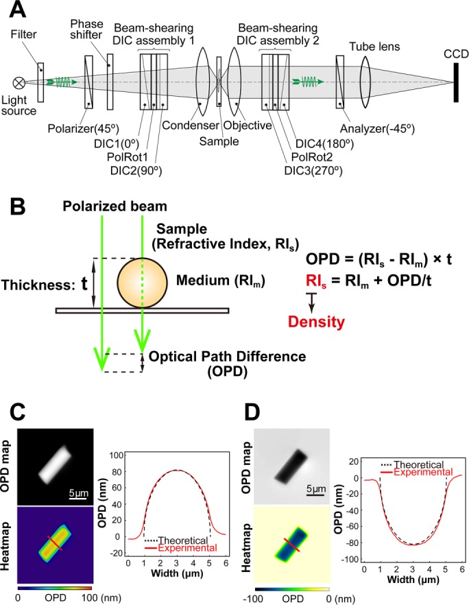
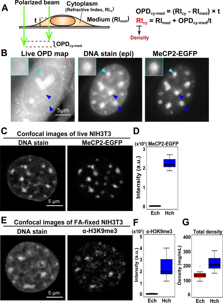
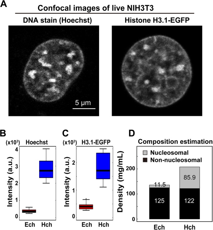
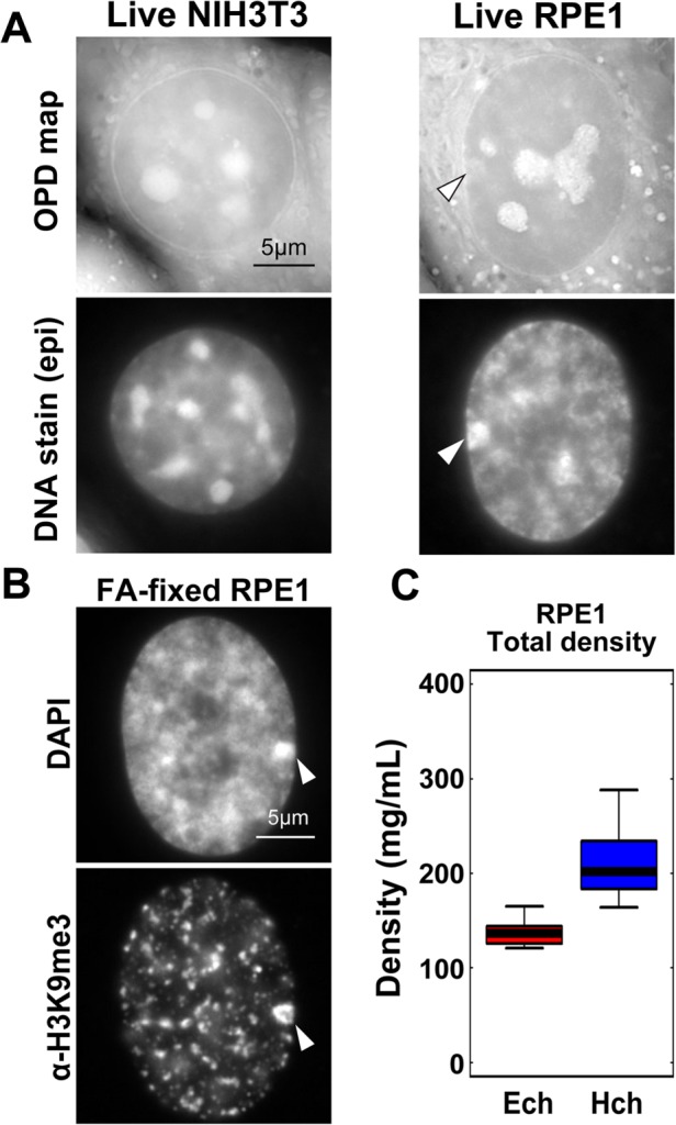
In eukaryotic cells, highly condensed inactive/silenced chromatin has long been called "heterochromatin." However, recent research suggests that such regions are in fact not fully transcriptionally silent and that there exists only a moderate access barrier to heterochromatin. To further investigate this issue, it is critical to elucidate the physical properties of heterochromatin such as its total density in live cells. Here, using orientation-independent differential interference contrast (OI-DIC) microscopy, which is capable of mapping optical path differences, we investigated the density of the total materials in pericentric foci, a representative heterochromatin model, in live mouse NIH3T3 cells. We demonstrated that the total density of heterochromatin (208 mg/ml) was only 1.53-fold higher than that of the surrounding euchromatic regions (136 mg/ml) while the DNA density of heterochromatin was 5.5- to 7.5-fold higher. We observed similar minor differences in density in typical facultative heterochromatin, the inactive human X chromosomes. This surprisingly small difference may be due to that nonnucleosomal materials (proteins/RNAs) (∼120 mg/ml) are dominant in both chromatin regions. Monte Carlo simulation suggested that nonnucleosomal materials contribute to creating a moderate access barrier to heterochromatin, allowing minimal protein access to functional regions. Our OI-DIC imaging offers new insight into the live cellular environments.
在真核细胞中,高度浓缩的无活性/沉默染色质长期以来一直被称为“异染色质”。然而,最近的研究表明,这些区域实际上并非完全转录沉默,并且对异染色质仅存在适度的进入屏障。为了进一步研究这个问题,阐明异染色质的物理性质,如活细胞中的总密度,至关重要。在这里,我们使用能够绘制光程差的非定向微分干涉对比(OI-DIC)显微镜,研究了活的小鼠NIH3T3细胞中着丝粒周围焦点(一种代表性的异染色质模型)中总物质的密度。我们证明,异染色质的总密度(208毫克/毫升)仅比周围常染色质区域(136毫克/毫升)高1.53倍,而异染色质的DNA密度高5.5至7.5倍。我们在典型的兼性异染色质(无活性的人类X染色体)中观察到了类似的微小密度差异。这种惊人的小差异可能是由于非核小体物质(蛋白质/RNA)(约120毫克/毫升)在两个染色质区域中占主导地位。蒙特卡罗模拟表明,非核小体物质有助于为异染色质创造适度的进入屏障,使蛋白质极少进入功能区域。我们的OI-DIC成像为活细胞环境提供了新的见解。