Department of Molecular, Cellular and Integrative Physiology, University of California at Los Angeles, Los Angeles, California, USA.
Department of Chemical and Systems Biology, Stanford University, Stanford, California, USA.
Sci Rep. 2017 Aug 23;7(1):9195. doi: 10.1038/s41598-017-09697-x.
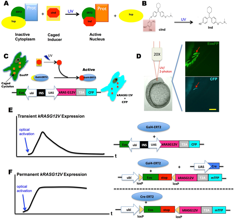
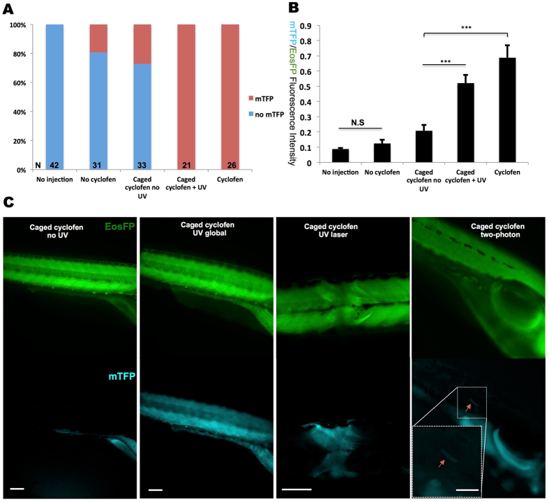
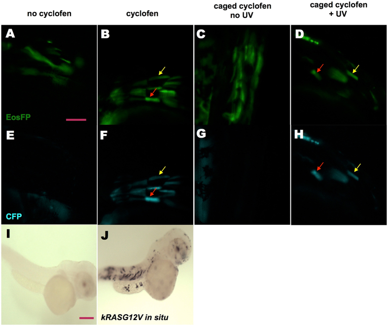
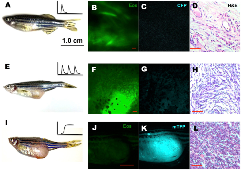
The zebrafish has become an increasingly popular and valuable cancer model over the past few decades. While most zebrafish cancer models are generated by expressing mammalian oncogenes under tissue-specific promoters, here we describe a method that allows for the precise optical control of oncogene expression in live zebrafish. We utilize this technique to transiently or constitutively activate a typical human oncogene, kRASG12V, in zebrafish embryos and investigate the developmental and tumorigenic phenotypes. We demonstrate the spatiotemporal control of oncogene expression in live zebrafish, and characterize the different tumorigenic probabilities when kRASG12V is expressed transiently or constitutively at different developmental stages. Moreover, we show that light can be used to activate oncogene expression in selected tissues and single cells without tissue-specific promoters. Our work presents a novel approach to initiate and study cancer in zebrafish, and the high spatiotemporal resolution of this method makes it a valuable tool for studying cancer initiation from single cells.
在过去的几十年里,斑马鱼已成为一种越来越受欢迎且极具价值的癌症模型。虽然大多数斑马鱼癌症模型是通过在组织特异性启动子下表达哺乳动物致癌基因来产生的,但在这里我们描述了一种可在活体斑马鱼中精确光学控制致癌基因表达的方法。我们利用该技术在斑马鱼胚胎中瞬时或持续激活典型的人类致癌基因 kRASG12V,并研究其发育和致瘤表型。我们证明了在活体斑马鱼中进行致癌基因表达的时空控制,并描述了当 kRASG12V 在不同发育阶段瞬时或持续表达时,其不同的致瘤概率。此外,我们还表明,可以在没有组织特异性启动子的情况下,使用光在选定的组织和单个细胞中激活致癌基因表达。我们的工作为在斑马鱼中引发和研究癌症提供了一种新方法,并且该方法具有较高的时空分辨率,使其成为从单细胞研究癌症起始的有价值的工具。