Apostolidis Leonidas, Schwarz Daniel, Xia Annie, Weiler Markus, Heckel Andreas, Godel Tim, Heiland Sabine, Schlemmer Heinz-Peter, Jäger Dirk, Bendszus Martin, Bäumer Philipp
Department of Medical Oncology, National Center for Tumor Diseases, Heidelberg, Germany.
Department of Neuroradiology, Heidelberg University Hospital, Heidelberg, Germany.
PLoS One. 2017 Aug 24;12(8):e0183845. doi: 10.1371/journal.pone.0183845. eCollection 2017.
To investigate in vivo morphological and functional correlates of oxaliplatin-induced peripheral neuropathy (OXA-PNP) by magnetic resonance neurography (MRN).
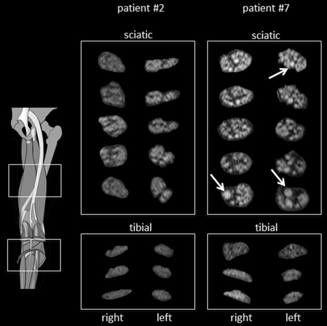
Twenty patients (7 female, 13 male, 58.9±10.0 years) with mild to moderate OXA-PNP and 20 matched controls (8 female, 12 male, 55.7±15.6 years) were prospectively enrolled. All patients underwent a detailed neurophysiological examination prior to neuroimaging. A standardized imaging protocol at 3.0 Tesla included the lumbosacral plexus and both sciatic nerves and their branches using T2-weighted fat-saturated sequences and diffusion tensor imaging. Quantitative assessment included volumetry of the dorsal root ganglia (DRG), sciatic nerve normalized T2 (nT2) signal and caliber, and fractional anisotropy (FA), mean diffusivity (MD), axial (AD) and radial diffusivity (RD). Additional qualitative evaluation of sciatic, peroneal, and tibial nerves evaluated the presence, degree, and distribution of nerve lesions.
DRG hypertrophy in OXA-PNP patients (207.3±47.7mm3 vs. 153.0±47.1mm3 in controls, p = 0.001) was found as significant morphological correlate of the sensory neuronopathy. In contrast, peripheral nerves only exhibited minor morphological alterations qualitatively. Quantitatively, sciatic nerve caliber (27.3±6.7mm2 vs. 27.4±7.4mm2, p = 0.80) and nT2 signal were not significantly changed in patients (1.32±0.22 vs. 1.22±0.26, p = 0.16). AD, RD, and MD showed a non-significant decrease in patients, while FA was unchanged.
OXA-PNP manifests with morphological and functional correlates that can be detected in vivo by MRN. We report hypertrophy of the DRG that stands in contrast to experimental and postmortem studies. DRG volume should be further investigated as a biomarker in other sensory peripheral neuropathies and ganglionopathies.
通过磁共振神经成像(MRN)研究奥沙利铂诱导的周围神经病变(OXA-PNP)的体内形态学和功能相关性。
前瞻性纳入20例轻度至中度OXA-PNP患者(7例女性,13例男性,年龄58.9±10.0岁)和20例匹配的对照者(8例女性,12例男性,年龄55.7±15.6岁)。所有患者在神经成像检查前均接受了详细的神经生理学检查。在3.0特斯拉的标准化成像方案包括使用T2加权脂肪抑制序列和扩散张量成像对腰骶丛、双侧坐骨神经及其分支进行成像。定量评估包括背根神经节(DRG)体积、坐骨神经标准化T2(nT2)信号和管径,以及分数各向异性(FA)、平均扩散率(MD)、轴向扩散率(AD)和径向扩散率(RD)。对坐骨神经、腓总神经和胫神经进行额外的定性评估,以评估神经病变的存在情况、程度和分布。
发现OXA-PNP患者的DRG肥大(207.3±47.7mm³,对照组为153.0±47.1mm³,p = 0.001)是感觉神经元病的显著形态学相关性表现。相比之下,周围神经仅表现出轻微的定性形态学改变。定量分析显示,患者的坐骨神经管径(27.3±6.7mm²,对照组为27.4±7.4mm²,p = 0.80)和nT2信号无显著变化(1.32±0.22,对照组为1.22±0.26,p = 0.16)。患者的AD、RD和MD呈非显著性降低,而FA无变化。
OXA-PNP表现出可通过MRN在体内检测到的形态学和功能相关性。我们报告了DRG肥大,这与实验研究和尸检研究结果相反。DRG体积应作为其他感觉性周围神经病变和神经节病变的生物标志物进行进一步研究。