• 文献检索
  • 文档翻译
  • 深度研究
  • 学术资讯
  • Suppr Zotero 插件Zotero 插件
  • 邀请有礼
  • 套餐&价格
  • 历史记录
应用&插件
Suppr Zotero 插件Zotero 插件浏览器插件Mac 客户端Windows 客户端微信小程序
定价
高级版会员购买积分包购买API积分包
服务
文献检索文档翻译深度研究API 文档MCP 服务
关于我们
关于 Suppr公司介绍联系我们用户协议隐私条款
关注我们

Suppr 超能文献

核心技术专利:CN118964589B侵权必究
粤ICP备2023148730 号-1Suppr @ 2026

文献检索

告别复杂PubMed语法,用中文像聊天一样搜索,搜遍4000万医学文献。AI智能推荐,让科研检索更轻松。

立即免费搜索

文件翻译

保留排版,准确专业,支持PDF/Word/PPT等文件格式,支持 12+语言互译。

免费翻译文档

深度研究

AI帮你快速写综述,25分钟生成高质量综述,智能提取关键信息,辅助科研写作。

立即免费体验

衰老过程中全脑结构变化的磁共振成像评估

MRI assessment of whole-brain structural changes in aging.

作者信息

Guo Hui, Siu William, D'Arcy Ryan Cn, Black Sandra E, Grajauskas Lukas A, Singh Sonia, Zhang Yunting, Rockwood Kenneth, Song Xiaowei

机构信息

Health Sciences and Innovation, ImageTech Laboratory, Fraser Health Authority, Surrey, BC, Canada.

Department of Diagnostic Imaging, Tianjin Medical University General Hospital, Tianjin, China.

出版信息

Clin Interv Aging. 2017 Aug 9;12:1251-1270. doi: 10.2147/CIA.S139515. eCollection 2017.

DOI:10.2147/CIA.S139515
PMID:28848333
原文链接:https://pmc.ncbi.nlm.nih.gov/articles/PMC5557118/
Abstract

PURPOSE

One of the central features of brain aging is the accumulation of multiple age-related structural changes, which occur heterogeneously in individuals and can have immediate or potential clinical consequences. Each of these deficits can coexist and interact, producing both independent and additive impacts on brain health. Many of the changes can be visualized using MRI. To collectively assess whole-brain structural changes, the MRI-based Brain Atrophy and Lesion Index (BALI) has been developed. In this study, we validate this whole-brain health assessment approach using several clinical MRI examinations.

MATERIALS AND METHODS

Data came from three independent studies: the Alzheimer's Disease Neuroimaging Initiative Phase II (n=950; women =47.9%; age =72.7±7.4 years); the National Alzheimer's Coordinating Center (n=722; women =55.1%; age =72.7±9.9 years); and the Tianjin Medical University General Hospital Research database on older adults (n=170; women =60.0%; age =62.9±9.3 years). The 3.0-Tesla MRI scans were evaluated using the BALI rating scheme on the basis of T1-weighted (T1WI), T2-weighted (T2WI), T2-weighted fluid-attenuated inversion recovery (T2-FLAIR), and T2*-weighted gradient-recalled echo (T2*GRE) images.

RESULTS

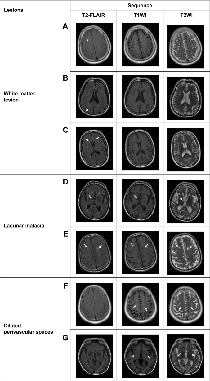
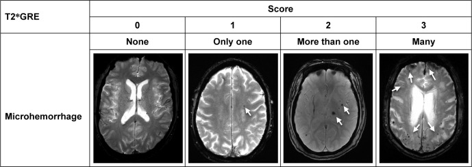
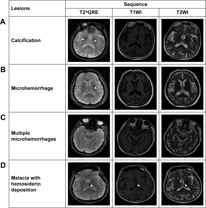
Atrophy and lesion changes were commonly seen in each MRI test. The BALI scores based on different sequences were highly correlated (Spearman >0.69; <0.00001). They were associated with age (>0.29; <0.00001) and differed by cognitive status (>26.48, <0.00001). T2-FLAIR revealed a greater level of periventricular (=29.09) and deep white matter (=26.65, <0.001) lesions than others, but missed revealing certain dilated perivascular spaces that were seen in T2WI (<0.001). Microhemorrhages occurred in 15.3% of the sample examined and were detected using only T2*GRE.

CONCLUSION

The T1WI- and T2WI-based BALI evaluations consistently identified the burden of aging and dementia-related decline of structural brain health. Inclusion of additional MRI tests increased lesion differentiation. Further research is to integrate MRI tests for a clinical tool to aid the diagnosis and intervention of brain aging.

摘要

目的

脑老化的核心特征之一是多种与年龄相关的结构变化的积累,这些变化在个体中异质性发生,可能产生直接或潜在的临床后果。这些缺陷中的每一个都可能共存并相互作用,对脑健康产生独立和累加的影响。许多变化可以通过磁共振成像(MRI)可视化。为了综合评估全脑结构变化,基于MRI的脑萎缩和病变指数(BALI)已被开发出来。在本研究中,我们使用几种临床MRI检查来验证这种全脑健康评估方法。

材料与方法

数据来自三项独立研究:阿尔茨海默病神经影像学倡议二期(n = 950;女性 = 47.9%;年龄 = 72.7±7.4岁);国家阿尔茨海默病协调中心(n = 722;女性 = 55.1%;年龄 = 72.7±9.9岁);以及天津医科大学总医院老年成人研究数据库(n = 170;女性 = 60.0%;年龄 = 62.9±9.3岁)。基于T1加权(T1WI)、T2加权(T2WI)、T2加权液体衰减反转恢复(T2-FLAIR)和T2加权梯度回波(T2GRE)图像,使用BALI评分方案对3.0特斯拉MRI扫描进行评估。

结果

在每次MRI检查中,萎缩和病变变化都很常见。基于不同序列的BALI评分高度相关(斯皮尔曼相关系数>0.69;P<0.00001)。它们与年龄相关(相关系数>0.29;P<0.00001),并且在认知状态方面存在差异(差异>26.48,P<0.00001)。T2-FLAIR显示出比其他序列更高水平的脑室周围病变(P = 29.09)和深部白质病变(P = 26.65,P<0.001),但遗漏了在T2WI中可见的某些扩张的血管周围间隙(P<0.001)。在所检查的样本中,15.3%出现微出血,且仅使用T2*GRE检测到。

结论

基于T1WI和T2WI的BALI评估一致地识别出与衰老和痴呆相关的脑结构健康下降的负担。纳入额外的MRI检查增加了病变的区分度。进一步的研究是将MRI检查整合为一种临床工具,以辅助脑老化的诊断和干预。

https://cdn.ncbi.nlm.nih.gov/pmc/blobs/9f97/5557118/49ded07b614e/cia-12-1251Fig3.jpg
https://cdn.ncbi.nlm.nih.gov/pmc/blobs/9f97/5557118/203dd1dfd4df/cia-12-1251Fig1.jpg
https://cdn.ncbi.nlm.nih.gov/pmc/blobs/9f97/5557118/35740eb1f656/cia-12-1251Fig2.jpg
https://cdn.ncbi.nlm.nih.gov/pmc/blobs/9f97/5557118/49ded07b614e/cia-12-1251Fig3.jpg
https://cdn.ncbi.nlm.nih.gov/pmc/blobs/9f97/5557118/203dd1dfd4df/cia-12-1251Fig1.jpg
https://cdn.ncbi.nlm.nih.gov/pmc/blobs/9f97/5557118/35740eb1f656/cia-12-1251Fig2.jpg
https://cdn.ncbi.nlm.nih.gov/pmc/blobs/9f97/5557118/49ded07b614e/cia-12-1251Fig3.jpg

相似文献

1
MRI assessment of whole-brain structural changes in aging.衰老过程中全脑结构变化的磁共振成像评估
Clin Interv Aging. 2017 Aug 9;12:1251-1270. doi: 10.2147/CIA.S139515. eCollection 2017.
2
Evaluation of common structural brain changes in aging and Alzheimer disease with the use of an MRI-based brain atrophy and lesion index: a comparison between T1WI and T2WI at 1.5T and 3T.利用基于磁共振成像的脑萎缩和病变指数评估衰老和阿尔茨海默病中常见的脑结构变化:1.5T和3T场强下T1加权成像与T2加权成像的比较
AJNR Am J Neuroradiol. 2014 Mar;35(3):504-12. doi: 10.3174/ajnr.A3709. Epub 2013 Aug 29.
3
An MRI brain atrophy and lesion index to assess the progression of structural changes in Alzheimer's disease, mild cognitive impairment, and normal aging: a follow-up study.阿尔茨海默病、轻度认知障碍和正常老化的结构变化进展评估的 MRI 脑萎缩和病变指数:一项随访研究。
J Alzheimers Dis. 2011;26 Suppl 3:359-67. doi: 10.3233/JAD-2011-0048.
4
Evaluation of whole brain health in aging and Alzheimer's disease: a standard procedure for scoring an MRI-based brain atrophy and lesion index.衰老和阿尔茨海默病中全脑健康的评估:基于MRI的脑萎缩和病变指数评分的标准程序。
J Alzheimers Dis. 2014;42(2):691-703. doi: 10.3233/JAD-140333.
5
MarkVCID cerebral small vessel consortium: II. Neuroimaging protocols.马克 VCID 脑小血管联盟:二、神经影像学协议。
Alzheimers Dement. 2021 Apr;17(4):716-725. doi: 10.1002/alz.12216. Epub 2021 Jan 21.
6
Combining structural brain changes improves the prediction of Alzheimer's disease and mild cognitive impairment.结合结构脑变化可提高阿尔茨海默病和轻度认知障碍的预测能力。
Dement Geriatr Cogn Disord. 2012;33(5):318-26. doi: 10.1159/000339364. Epub 2012 Jul 2.
7
Dynamics of brain structure and cognitive function in the Alzheimer's disease neuroimaging initiative.阿尔茨海默病神经影像学倡议中的大脑结构和认知功能的动态变化。
J Neurol Neurosurg Psychiatry. 2013 Jan;84(1):71-8. doi: 10.1136/jnnp-2012-303579. Epub 2012 Nov 2.
8
Feasibility of Brain Atrophy Measurement in Clinical Routine without Prior Standardization of the MRI Protocol: Results from MS-MRIUS, a Longitudinal Observational, Multicenter Real-World Outcome Study in Patients with Relapsing-Remitting MS.临床常规中不进行 MRI 方案标准化而进行脑萎缩测量的可行性:来自 MS-MRIUS 的结果,这是一项在复发缓解型多发性硬化症患者中进行的纵向观察性、多中心真实世界结局研究。
AJNR Am J Neuroradiol. 2018 Feb;39(2):289-295. doi: 10.3174/ajnr.A5442. Epub 2017 Nov 23.
9
Toward MRI-based whole-brain health assessment: The brain atrophy and lesion index (BALI).迈向基于磁共振成像的全脑健康评估:脑萎缩与病变指数(BALI)。
Aging Med (Milton). 2018 Apr 26;1(1):55-63. doi: 10.1002/agm2.12014. eCollection 2018 Jun.
10
The reliability of magnetic resonance imaging in traumatic brain injury lesion detection.磁共振成像在创伤性脑损伤病变检测中的可靠性。
Brain Inj. 2012;26(12):1439-50. doi: 10.3109/02699052.2012.694563. Epub 2012 Jun 25.

引用本文的文献

1
Antiageing strategy for neurodegenerative diseases: from mechanisms to clinical advances.神经退行性疾病的抗衰老策略:从机制到临床进展
Signal Transduct Target Ther. 2025 Mar 10;10(1):76. doi: 10.1038/s41392-025-02145-7.
2
Factors associated with attitudes toward research MRI in older Asian Americans.与年长亚裔美国人对研究性磁共振成像态度相关的因素。
Alzheimers Dement (N Y). 2024 Feb 9;10(1):e12449. doi: 10.1002/trc2.12449. eCollection 2024 Jan-Mar.
3
Arteriolar neuropathology in cerebral microvascular disease.脑微血管病的小动脉神经病理学。

本文引用的文献

1
Radiological-Pathological Correlation in Alzheimer's Disease: Systematic Review of Antemortem Magnetic Resonance Imaging Findings.阿尔茨海默病的放射学与病理学相关性:生前磁共振成像结果的系统综述
J Alzheimers Dis. 2017;57(2):575-601. doi: 10.3233/JAD-161028.
2
Early role of vascular dysregulation on late-onset Alzheimer's disease based on multifactorial data-driven analysis.基于多因素数据驱动分析的血管调节异常在迟发性阿尔茨海默病中的早期作用。
Nat Commun. 2016 Jun 21;7:11934. doi: 10.1038/ncomms11934.
3
Density abnormalities in normal-appearing gray matter in the middle-aged brain with white matter hyperintense lesions: a DARTEL-enhanced voxel-based morphometry study.
Neuropathol Appl Neurobiol. 2023 Feb;49(1):e12875. doi: 10.1111/nan.12875.
4
Aging Brains Degrade Driving Safety Performances of the Healthy Elderly.衰老的大脑会降低健康老年人的驾驶安全表现。
Front Aging Neurosci. 2022 Jan 25;13:783717. doi: 10.3389/fnagi.2021.783717. eCollection 2021.
5
3D hemisphere-based convolutional neural network for whole-brain MRI segmentation.基于 3D 半球的卷积神经网络进行全脑 MRI 分割。
Comput Med Imaging Graph. 2022 Jan;95:102000. doi: 10.1016/j.compmedimag.2021.102000. Epub 2021 Oct 30.
6
Assessment of structural brain changes in patients with type 2 diabetes mellitus using the MRI-based brain atrophy and lesion index.使用基于MRI的脑萎缩和病变指数评估2型糖尿病患者的脑结构变化。
Neural Regen Res. 2022 Mar;17(3):618-624. doi: 10.4103/1673-5374.320996.
7
Normal-sized basal ganglia perivascular space related to motor phenotype in Parkinson freezers.正常大小基底节血管周围空间与帕金森冻结步态患者的运动表型相关。
Aging (Albany NY). 2021 Jul 27;13(14):18912-18923. doi: 10.18632/aging.203343.
8
Searching for female reproductive aging and longevity biomarkers.寻找女性生殖衰老和长寿的生物标志物。
Aging (Albany NY). 2021 Jun 22;13(12):16873-16894. doi: 10.18632/aging.203206.
9
Apolipoprotein E ε4 Allele-Based Differences in Brain Volumes Are Largely Uniform Across Late Middle Aged and Older Hispanic/Latino- and Non-Hispanic/Latino Whites Without Dementia.基于载脂蛋白Eε4等位基因的脑容量差异在无痴呆症的中老年西班牙裔/拉丁裔和非西班牙裔/拉丁裔白人中基本一致。
Front Aging Neurosci. 2021 Feb 26;13:627322. doi: 10.3389/fnagi.2021.627322. eCollection 2021.
10
Age-Related Alterations in Brain Perfusion, Venous Oxygenation, and Oxygen Metabolic Rate of Mice: A 17-Month Longitudinal MRI Study.小鼠脑灌注、静脉氧合及氧代谢率的年龄相关性改变:一项为期17个月的纵向MRI研究
Front Neurol. 2020 Jun 12;11:559. doi: 10.3389/fneur.2020.00559. eCollection 2020.
中年大脑中伴有白质高信号病变的正常灰质密度异常:一项基于DARTEL增强体素的形态学研究
Clin Interv Aging. 2016 May 12;11:615-22. doi: 10.2147/CIA.S98409. eCollection 2016.
4
Imaging the Perivascular Space as a Potential Biomarker of Neurovascular and Neurodegenerative Diseases.将血管周围间隙成像作为神经血管和神经退行性疾病的潜在生物标志物
Cell Mol Neurobiol. 2016 Mar;36(2):289-99. doi: 10.1007/s10571-016-0343-6. Epub 2016 Mar 18.
5
Visual Assessment of Age-Related White Matter Hyperintensities Using FLAIR Images at 3 T: Inter- and Intra-Rater Agreement.在3T场强下使用液体衰减反转恢复(FLAIR)图像对年龄相关性脑白质高信号进行视觉评估:评分者间和评分者内一致性
Neurodegener Dis. 2016;16(3-4):279-83. doi: 10.1159/000441420. Epub 2015 Dec 9.
6
Topography of Cortical Microbleeds in Alzheimer's Disease with and without Cerebral Amyloid Angiopathy: A Post-Mortem 7.0-Tesla MRI Study.伴和不伴脑淀粉样血管病的阿尔茨海默病患者皮质微出血的分布情况:一项7.0特斯拉尸检MRI研究
Aging Dis. 2015 Nov 17;6(6):437-43. doi: 10.14336/AD.2015.0429. eCollection 2015 Nov.
7
Cerebral small vessel disease and Alzheimer's disease.脑小血管病与阿尔茨海默病
Clin Interv Aging. 2015 Oct 23;10:1695-704. doi: 10.2147/CIA.S90871. eCollection 2015.
8
White matter hyperintensities, cognitive impairment and dementia: an update.脑白质高信号、认知障碍与痴呆:最新进展。
Nat Rev Neurol. 2015 Mar;11(3):157-65. doi: 10.1038/nrneurol.2015.10. Epub 2015 Feb 17.
9
Pathological correlates of white matter hyperintensities on magnetic resonance imaging.磁共振成像上白质高信号的病理相关性
Dement Geriatr Cogn Disord. 2015;39(1-2):92-104. doi: 10.1159/000366411. Epub 2014 Nov 8.
10
A comprehensive visual rating scale of brain magnetic resonance imaging: application in elderly subjects with Alzheimer's disease, mild cognitive impairment, and normal cognition.脑磁共振成像的综合视觉评分量表:在患有阿尔茨海默病、轻度认知障碍和认知正常的老年受试者中的应用。
J Alzheimers Dis. 2015;44(3):1023-34. doi: 10.3233/JAD-142088.