• 文献检索
  • 文档翻译
  • 深度研究
  • 学术资讯
  • Suppr Zotero 插件Zotero 插件
  • 邀请有礼
  • 套餐&价格
  • 历史记录
应用&插件
Suppr Zotero 插件Zotero 插件浏览器插件Mac 客户端Windows 客户端微信小程序
定价
高级版会员购买积分包购买API积分包
服务
文献检索文档翻译深度研究API 文档MCP 服务
关于我们
关于 Suppr公司介绍联系我们用户协议隐私条款
关注我们

Suppr 超能文献

核心技术专利:CN118964589B侵权必究
粤ICP备2023148730 号-1Suppr @ 2026

文献检索

告别复杂PubMed语法,用中文像聊天一样搜索,搜遍4000万医学文献。AI智能推荐,让科研检索更轻松。

立即免费搜索

文件翻译

保留排版,准确专业,支持PDF/Word/PPT等文件格式,支持 12+语言互译。

免费翻译文档

深度研究

AI帮你快速写综述,25分钟生成高质量综述,智能提取关键信息,辅助科研写作。

立即免费体验

三种不同光学相干断层扫描血管造影仪测量的中心凹无血管区面积的可重复性和差异。

Reproducibility and differences in area of foveal avascular zone measured by three different optical coherence tomographic angiography instruments.

机构信息

Department of Ophthalmology, Kagoshima University Graduate School of Medical and Dental Sciences, Kagoshima, Japan.

出版信息

Sci Rep. 2017 Aug 29;7(1):9853. doi: 10.1038/s41598-017-09255-5.

DOI:10.1038/s41598-017-09255-5
PMID:28851930
原文链接:https://pmc.ncbi.nlm.nih.gov/articles/PMC5575252/
Abstract

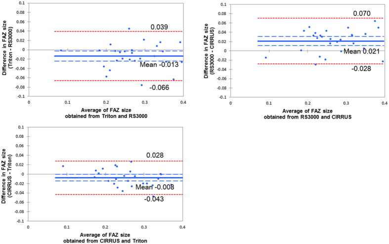
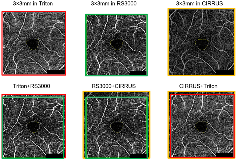
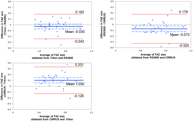
This study was performed to compare the area of the foveal avascular zone (FAZ-area) obtained by three optical coherence tomography angiography (OCTA) instruments. This was a cross-sectional, non-interventional study of twenty-seven healthy right eyes. The superficial and deep FAZ-area was measured manually with three OCTA instruments: Triton (Topcon), RS3000 (Nidek), and CIRRUS (Zeiss). The intra-rater, inter-rater, and inter-instrument correlation coefficients (CC) were assessed. The intra-rater and inter-rater CC were significantly high for the superficial and deep FAZ-areas (P < 0.001). The inter-instrument CC (95% confidence interval) for the superficial FAZ-area was 0.920 (0.803-0.965) for Triton vs RS3000, 0.899 (0.575-0.965) for RS3000 vs CIRRUS, and was 0.963 (0.913-0.983) for CIRRUS vs Triton (P < 0.001). For the deep FAZ-area, the inter-instrument CC was 0.813 (0.633-0.910) for Triton vs RS3000, 0.694 (0.369-0.857) for RS3000 vs CIRRUS, and 0.679 (0.153-0.872) for CIRRUS vs Triton (P < 0.001). The superficial FAZ-area (mm) was 0.264 ± 0.071 with Triton, 0.278 ± 0.072 with RS3000 and 0.257 ± 0.066 with CIRRUS. For deep FAZ-area, it was 0.617 ± 0.175 with Triton, 0.646 ± 0.178 with RS3000 and 0.719 ± 0.175 with CIRRUS. The FAZ-area from these instruments was clinically interchangeable. However, the absolute values of FAZ-area are significantly different among them. These differences must be considered in comparing the FAZ-areas from different OCTA instruments.

摘要

本研究旨在比较三种光学相干断层扫描血管造影(OCTA)仪器测量的中心凹无血管区(FAZ)面积。这是一项对 27 只健康右眼进行的横断面、非干预性研究。使用三种 OCTA 仪器(Triton、RS3000 和 Cirrus)手动测量浅层和深层 FAZ 区域。评估了内部观察者、外部观察者和仪器间的相关系数(CC)。浅层和深层 FAZ 区域的内部观察者和外部观察者 CC 显著较高(P<0.001)。浅层 FAZ 区域的仪器间 CC(95%置信区间)为 Triton 与 RS3000 之间为 0.920(0.803-0.965),RS3000 与 Cirrus 之间为 0.899(0.575-0.965),Cirrus 与 Triton 之间为 0.963(0.913-0.983)(P<0.001)。对于深层 FAZ 区域,仪器间 CC 为 Triton 与 RS3000 之间为 0.813(0.633-0.910),RS3000 与 Cirrus 之间为 0.694(0.369-0.857),Cirrus 与 Triton 之间为 0.679(0.153-0.872)(P<0.001)。浅层 FAZ 区域(mm)用 Triton 为 0.264±0.071,用 RS3000 为 0.278±0.072,用 Cirrus 为 0.257±0.066。深层 FAZ 区域用 Triton 为 0.617±0.175,用 RS3000 为 0.646±0.178,用 Cirrus 为 0.719±0.175。这些仪器的 FAZ 区域在临床上可互换。然而,它们之间 FAZ 区域的绝对值有显著差异。在比较不同 OCTA 仪器的 FAZ 区域时,必须考虑这些差异。

https://cdn.ncbi.nlm.nih.gov/pmc/blobs/2364/5575252/b76119b0f12f/41598_2017_9255_Fig3_HTML.jpg
https://cdn.ncbi.nlm.nih.gov/pmc/blobs/2364/5575252/f0c63a14888b/41598_2017_9255_Fig1_HTML.jpg
https://cdn.ncbi.nlm.nih.gov/pmc/blobs/2364/5575252/e01e16c376be/41598_2017_9255_Fig2_HTML.jpg
https://cdn.ncbi.nlm.nih.gov/pmc/blobs/2364/5575252/b76119b0f12f/41598_2017_9255_Fig3_HTML.jpg
https://cdn.ncbi.nlm.nih.gov/pmc/blobs/2364/5575252/f0c63a14888b/41598_2017_9255_Fig1_HTML.jpg
https://cdn.ncbi.nlm.nih.gov/pmc/blobs/2364/5575252/e01e16c376be/41598_2017_9255_Fig2_HTML.jpg
https://cdn.ncbi.nlm.nih.gov/pmc/blobs/2364/5575252/b76119b0f12f/41598_2017_9255_Fig3_HTML.jpg

相似文献

1
Reproducibility and differences in area of foveal avascular zone measured by three different optical coherence tomographic angiography instruments.三种不同光学相干断层扫描血管造影仪测量的中心凹无血管区面积的可重复性和差异。
Sci Rep. 2017 Aug 29;7(1):9853. doi: 10.1038/s41598-017-09255-5.
2
Comparative Evaluation of Foveal Avascular Zone on Two Optical Coherence Tomography Angiography Devices.两种光学相干断层扫描血管造影设备对黄斑无血管区的比较评估
Optom Vis Sci. 2018 Jul;95(7):602-607. doi: 10.1097/OPX.0000000000001238.
3
Reliability of foveal avascular zone metrics automatically measured by Cirrus optical coherence tomography angiography in healthy subjects.健康受试者中 Cirrus 光学相干断层扫描血管造影术自动测量的中心凹无血管区指标的可靠性。
Int Ophthalmol. 2020 Mar;40(3):763-773. doi: 10.1007/s10792-019-01238-x. Epub 2019 Dec 2.
4
Improved Automated Foveal Avascular Zone Measurement in Cirrus Optical Coherence Tomography Angiography Using the Level Sets Macro.利用水平集宏在 Cirrus 光学相干断层扫描血管造影中进行改进的自动黄斑无血管区测量。
Transl Vis Sci Technol. 2020 Nov 13;9(12):20. doi: 10.1167/tvst.9.12.20. eCollection 2020 Nov.
5
VARIABILITY IN FOVEAL AVASCULAR ZONE AND CAPILLARY DENSITY USING OPTICAL COHERENCE TOMOGRAPHY ANGIOGRAPHY MACHINES IN HEALTHY EYES.使用光学相干断层扫描血管造影机检测健康眼睛的黄斑中心凹血管区和毛细血管密度的变异性
Retina. 2017 Nov;37(11):2102-2111. doi: 10.1097/IAE.0000000000001458.
6
REPRODUCIBILITY AND RELIABILITY OF OPTICAL COHERENCE TOMOGRAPHY ANGIOGRAPHY FOR FOVEAL AVASCULAR ZONE EVALUATION AND MEASUREMENT IN DIFFERENT SETTINGS.不同环境下光学相干断层扫描血管造影术对黄斑中心凹血管区评估和测量的可重复性及可靠性
Retina. 2017 Sep;37(9):1636-1641. doi: 10.1097/IAE.0000000000001426.
7
Objective evaluation of size and shape of superficial foveal avascular zone in normal subjects by optical coherence tomography angiography.应用光相干断层扫描血管造影术客观评价正常人群浅层中心凹无血管区的大小和形态。
Sci Rep. 2018 Jul 4;8(1):10143. doi: 10.1038/s41598-018-28530-7.
8
Interocular symmetry of the foveal avascular zone area in healthy eyes: a swept-source optical coherence tomography angiography study.健康眼中黄斑区无血管区面积的双眼对称性:扫频源光学相干断层血管造影研究。
Jpn J Ophthalmol. 2020 Mar;64(2):171-179. doi: 10.1007/s10384-020-00719-2. Epub 2020 Feb 3.
9
Reproducibility of Foveal Avascular Zone and Superficial Macular Retinal Vasculature Measurements in Healthy Eyes Determined by Two Different Scanning Protocols of Optical Coherence Tomography Angiography.两种不同的光学相干断层扫描血管造影扫描方案在健康眼中对中心凹无血管区和浅层视网膜脉络膜血管测量的重复性。
Ophthalmic Res. 2020;63(3):244-251. doi: 10.1159/000503071. Epub 2019 Oct 16.
10
Different effect of media opacity on automated and manual measurement of foveal avascular zone of optical coherence tomography angiographies.不同的媒介不透明度对光学相干断层扫描血管造影的自动和手动黄斑无血管区测量的影响不同。
Br J Ophthalmol. 2021 Jun;105(6):812-818. doi: 10.1136/bjophthalmol-2019-315780. Epub 2020 Jul 23.

引用本文的文献

1
Vascular Perfusion Variability in Diabetic Retinopathy: A Sequential Interscan Optical Coherence Tomography Angiography Assessment.糖尿病视网膜病变中的血管灌注变异性:序列扫描间光学相干断层扫描血管造影评估
J Clin Med. 2025 Mar 28;14(7):2312. doi: 10.3390/jcm14072312.
2
Association of Microvasculature Changes with Visual Outcomes in Idiopathic Epiretinal Membrane Surgery: A Clinical Trial.特发性视网膜前膜手术中微血管变化与视觉预后的关联:一项临床试验
J Clin Med. 2024 Aug 13;13(16):4748. doi: 10.3390/jcm13164748.
3
Choriocapillaris and Retinal Vascular Alterations in Presymptomatic Alzheimer's Disease.

本文引用的文献

1
VARIABILITY IN FOVEAL AVASCULAR ZONE AND CAPILLARY DENSITY USING OPTICAL COHERENCE TOMOGRAPHY ANGIOGRAPHY MACHINES IN HEALTHY EYES.使用光学相干断层扫描血管造影机检测健康眼睛的黄斑中心凹血管区和毛细血管密度的变异性
Retina. 2017 Nov;37(11):2102-2111. doi: 10.1097/IAE.0000000000001458.
2
Retinal Capillary Density and Foveal Avascular Zone Area Are Age-Dependent: Quantitative Analysis Using Optical Coherence Tomography Angiography.视网膜毛细血管密度和黄斑无血管区面积与年龄相关:使用光学相干断层扫描血管造影术的定量分析。
Invest Ophthalmol Vis Sci. 2016 Oct 1;57(13):5780-5787. doi: 10.1167/iovs.16-20045.
3
Visual Acuity Is Correlated with the Area of the Foveal Avascular Zone in Diabetic Retinopathy and Retinal Vein Occlusion.
早发性阿尔茨海默病的脉络膜毛细血管和视网膜血管改变。
Invest Ophthalmol Vis Sci. 2024 Jan 2;65(1):47. doi: 10.1167/iovs.65.1.47.
4
Repeatability and reproducibility of quantitative OCT angiography measurements from table-top and portable Flex Spectralis devices.桌面式和便携式 Flex Spectralis 设备定量 OCT 血管造影测量的重复性和可再现性。
Graefes Arch Clin Exp Ophthalmol. 2024 Jun;262(6):1785-1793. doi: 10.1007/s00417-023-06351-3. Epub 2024 Jan 25.
5
Application of ImageJ in Optical Coherence Tomography Angiography (OCT-A): A Literature Review.ImageJ在光学相干断层扫描血管造影(OCT-A)中的应用:文献综述。
J Ophthalmol. 2023 Nov 22;2023:9479183. doi: 10.1155/2023/9479183. eCollection 2023.
6
The Effect of Circle of Willis Morphology on Retinal Blood Flow in Patients with Carotid Stenosis Measured by Optical Coherence Tomography Angiography.利用光学相干断层扫描血管造影术测量 Willis 环形态对颈动脉狭窄患者视网膜血流的影响。
J Clin Med. 2023 Aug 16;12(16):5335. doi: 10.3390/jcm12165335.
7
A standardized method to quantitatively analyze optical coherence tomography angiography images of the macular and peripapillary vessels.一种定量分析黄斑和视乳头周围血管光学相干断层扫描血管造影图像的标准化方法。
Int J Retina Vitreous. 2022 Oct 15;8(1):75. doi: 10.1186/s40942-022-00426-9.
8
Change of intraocular blood flow during treatment for thyroid eye disease.甲状腺眼病治疗期间眼内血流的变化。
Taiwan J Ophthalmol. 2022 Feb 28;12(1):97-100. doi: 10.4103/tjo.tjo_2_22. eCollection 2022 Jan-Mar.
9
Quantitative analysis of early retinal vascular changes in type 2 diabetic patients without clinical retinopathy by optical coherence tomography angiography.应用光相干断层扫描血管造影术定量分析 2 型糖尿病无临床视网膜病变患者的早期视网膜血管变化。
Int Ophthalmol. 2022 Feb;42(2):367-375. doi: 10.1007/s10792-022-02230-8. Epub 2022 Jan 31.
10
Normative data on the foveal avascular zone in a young healthy Irish population using optical coherence tomography angiography.利用光学相干断层扫描血管造影术获得的爱尔兰年轻健康人群黄斑中心无血管区的正常值数据。
Eur J Ophthalmol. 2022 Sep;32(5):2824-2832. doi: 10.1177/11206721211073446. Epub 2022 Jan 10.
视力与糖尿病视网膜病变和视网膜静脉阻塞的中心无血管区面积相关。
Ophthalmology. 2016 Nov;123(11):2352-2367. doi: 10.1016/j.ophtha.2016.07.008. Epub 2016 Aug 11.
4
Swept-Source OCT Angiography Imaging of the Foveal Avascular Zone and Macular Capillary Network Density in Diabetic Retinopathy.糖尿病视网膜病变中黄斑无血管区和黄斑毛细血管网络密度的扫频源光学相干断层扫描血管造影成像
Invest Ophthalmol Vis Sci. 2016 Jul 1;57(8):3907-13. doi: 10.1167/iovs.16-19570.
5
Retinal Capillary Network and Foveal Avascular Zone in Eyes with Vein Occlusion and Fellow Eyes Analyzed With Optical Coherence Tomography Angiography.光学相干断层扫描血管造影分析静脉阻塞患眼及对侧眼的视网膜毛细血管网络和黄斑无血管区
Invest Ophthalmol Vis Sci. 2016 Jul 1;57(9):OCT486-94. doi: 10.1167/iovs.15-18907.
6
Optical Coherence Tomography Angiography Evaluation of the Parafoveal Vasculature and Its Relationship With Ocular Factors.黄斑旁血管系统的光学相干断层扫描血管造影评估及其与眼部因素的关系。
Invest Ophthalmol Vis Sci. 2016 Jul 1;57(9):OCT224-34. doi: 10.1167/iovs.15-18869.
7
Optical Coherence Tomography Angiography in Retinal Vein Occlusion: Evaluation of Superficial and Deep Capillary Plexa.视网膜静脉阻塞中的光学相干断层扫描血管造影:浅层和深层毛细血管丛的评估
Am J Ophthalmol. 2016 Jan;161:160-71.e1-2. doi: 10.1016/j.ajo.2015.10.008. Epub 2015 Oct 23.
8
DETECTION OF MICROVASCULAR CHANGES IN EYES OF PATIENTS WITH DIABETES BUT NOT CLINICAL DIABETIC RETINOPATHY USING OPTICAL COHERENCE TOMOGRAPHY ANGIOGRAPHY.利用光学相干断层扫描血管造影术检测糖尿病患者但无临床糖尿病视网膜病变患者眼部的微血管变化
Retina. 2015 Nov;35(11):2364-70. doi: 10.1097/IAE.0000000000000882.
9
ENLARGEMENT OF FOVEAL AVASCULAR ZONE IN DIABETIC EYES EVALUATED BY EN FACE OPTICAL COHERENCE TOMOGRAPHY ANGIOGRAPHY.通过正面光学相干断层扫描血管造影评估糖尿病患者眼部黄斑血管区扩大情况。
Retina. 2015 Nov;35(11):2377-83. doi: 10.1097/IAE.0000000000000849.
10
OPTICAL COHERENCE TOMOGRAPHY ANGIOGRAPHY OF RETINAL VENOUS OCCLUSION.视网膜静脉阻塞的光学相干断层扫描血管造影
Retina. 2015 Nov;35(11):2323-31. doi: 10.1097/IAE.0000000000000811.