Kim Jong Hun, Kim Eun Young, Jin Gong Yong, Choi Jong Bum
Department of Thoracic and Cardiovascular Surgery, Chonbuk National University Medical School, Research Institute of Clinical Medicine of Chonbuk National University, Biomedical Research Institute of Chonbuk National University Hospital, Jeonju 54907, Korea.
Department of Radiology, Research Institute of Clinical Medicine of Chonbuk National University, Biomedical Research Institute of Chonbuk National University Hospital, Institute for Medical Sciences of Chonbuk National University Medical School, Jeonju 54907, Korea.
Korean J Radiol. 2017 Sep-Oct;18(5):773-785. doi: 10.3348/kjr.2017.18.5.773. Epub 2017 Jul 17.
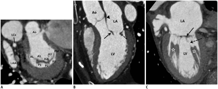
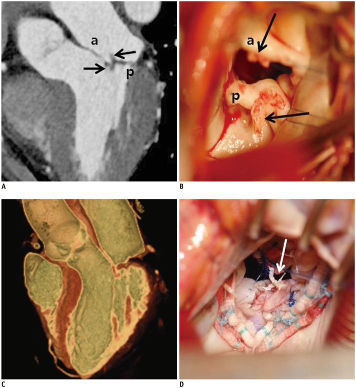
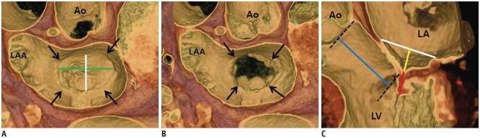
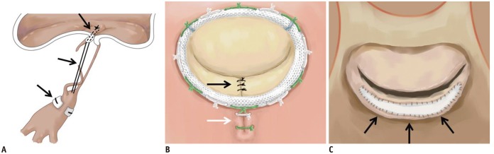
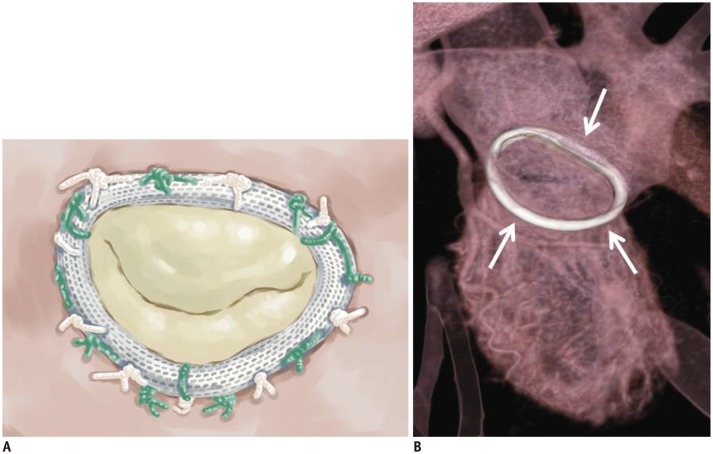
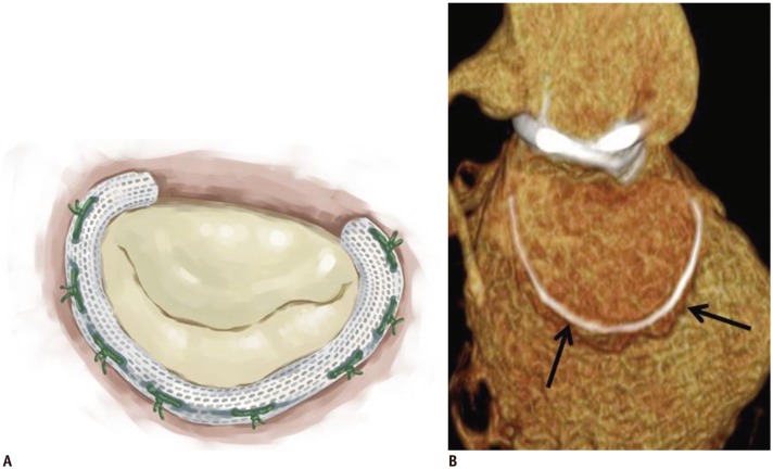
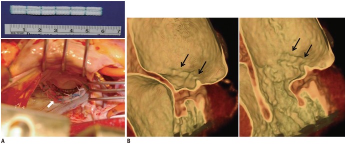
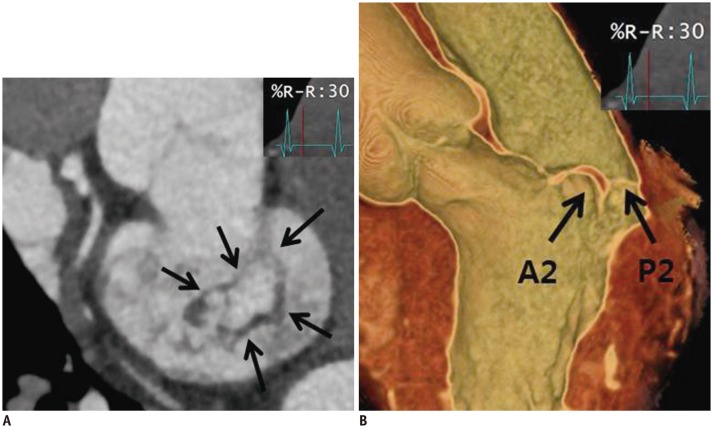
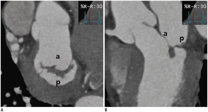
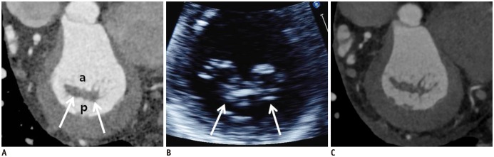
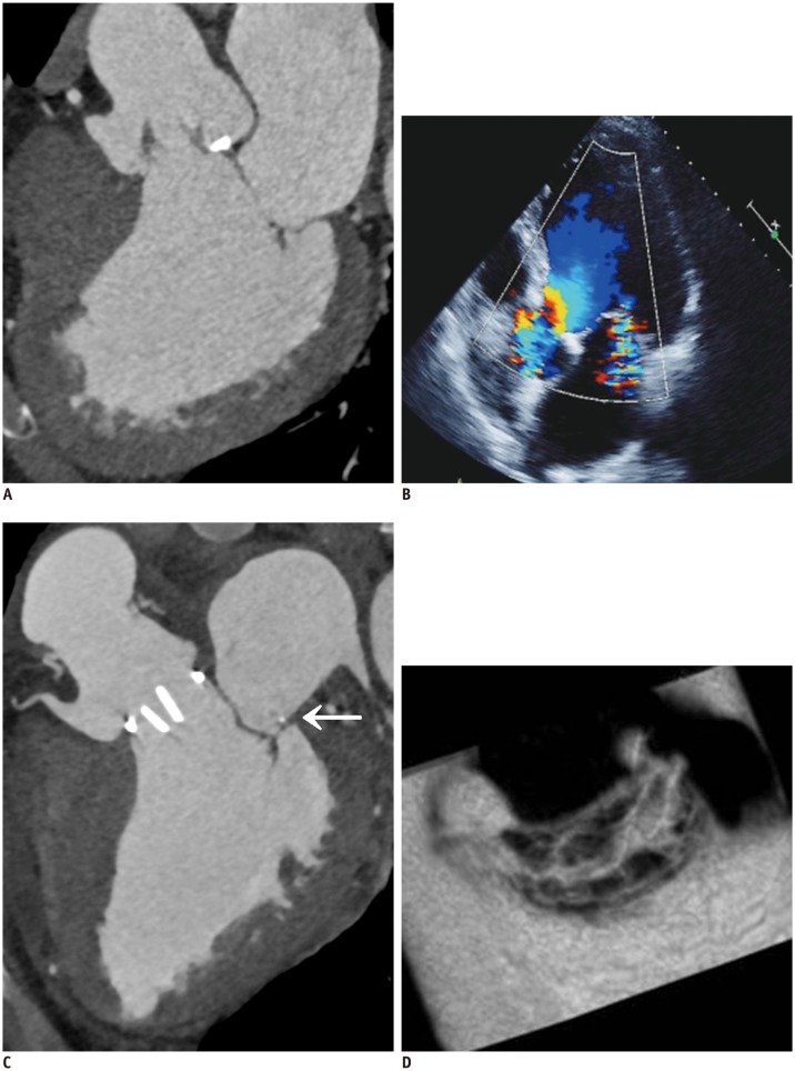
The role of cardiac computed tomography (CT) for evaluating the mitral valve (MV) has been limited since echocardiography is the main method of evaluation. However, recent advances in cardiac CT have enable detailed evaluation of the anatomy and geometry of the MV. We describe assessments of the anatomy and coaptation geometric parameters of normal MVs, and also review repair of diseased/damaged MV. We also discuss pre- and post-surgical imaging of MV pathology using cardiac CT and various CT images. We found that cardiac CT could be used as an alternative imaging modality to echocardiography for pre-operative MV evaluation and to predict clinical outcomes following repair.
由于超声心动图是评估二尖瓣(MV)的主要方法,心脏计算机断层扫描(CT)在评估二尖瓣方面的作用一直有限。然而,心脏CT的最新进展已能够对二尖瓣的解剖结构和几何形态进行详细评估。我们描述了正常二尖瓣的解剖结构和瓣叶对合几何参数的评估,同时也回顾了病变/受损二尖瓣的修复情况。我们还讨论了使用心脏CT和各种CT图像对二尖瓣病变进行手术前后的成像。我们发现,心脏CT可作为超声心动图的替代成像方式,用于二尖瓣术前评估以及预测修复后的临床结果。