• 文献检索
  • 文档翻译
  • 深度研究
  • 学术资讯
  • Suppr Zotero 插件Zotero 插件
  • 邀请有礼
  • 套餐&价格
  • 历史记录
应用&插件
Suppr Zotero 插件Zotero 插件浏览器插件Mac 客户端Windows 客户端微信小程序
定价
高级版会员购买积分包购买API积分包
服务
文献检索文档翻译深度研究API 文档MCP 服务
关于我们
关于 Suppr公司介绍联系我们用户协议隐私条款
关注我们

Suppr 超能文献

核心技术专利:CN118964589B侵权必究
粤ICP备2023148730 号-1Suppr @ 2026

文献检索

告别复杂PubMed语法,用中文像聊天一样搜索,搜遍4000万医学文献。AI智能推荐,让科研检索更轻松。

立即免费搜索

文件翻译

保留排版,准确专业,支持PDF/Word/PPT等文件格式,支持 12+语言互译。

免费翻译文档

深度研究

AI帮你快速写综述,25分钟生成高质量综述,智能提取关键信息,辅助科研写作。

立即免费体验

[培养的角膜缘上皮细胞用于角膜缘干细胞缺乏症的眼表重建:一年随访结果]

[Ocular Surface Reconstruction with Cultivated Limbal Epithelial Cells in Limbal Stem Cell Deficiency: One-year Follow-up Results].

作者信息

Durak İsmet, Selver Özlem Barut, Erdal Esra, Kunter İmge, Söylemezoğlu Zeynep Özbek, Wolosin Jose Mario

机构信息

Dokuz Eylül Üniversitesi Tıp Fakültesi, Göz Hastalıkları Anabilim Dalı İzmir, Türkiye.

Buca Seyfi Demirsoy Devlet Hastanesi, Göz Hastalıkları Birimi, İzmir, Türkiye.

出版信息

Turk Oftalmol Derg. 2012 May;42(3):172-176. doi: 10.4274/tjo.42.66375.

DOI:10.4274/tjo.42.66375
PMID:28868532
原文链接:https://pmc.ncbi.nlm.nih.gov/articles/PMC5576176/
Abstract

PURPOSE

To evaluate the 1-year follow-up results of cultivated limbal epithelial cell (CLEC) transplantation in unilateral limbal stem cell deficiency (LSCD).

MATERIAL AND METHOD

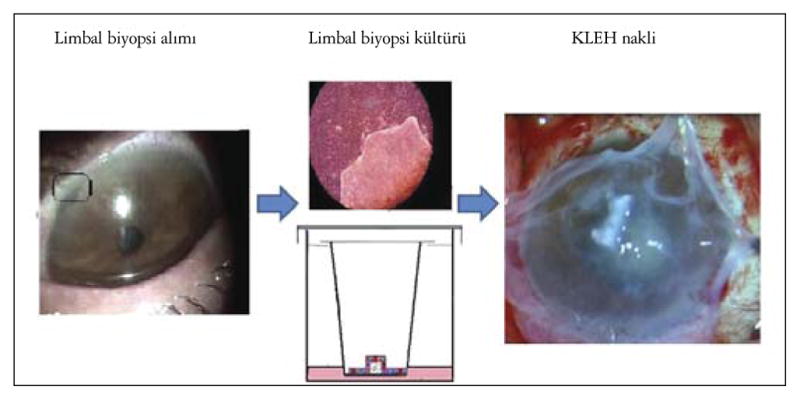
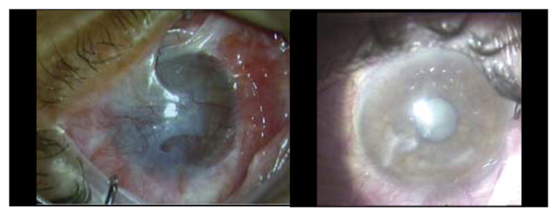
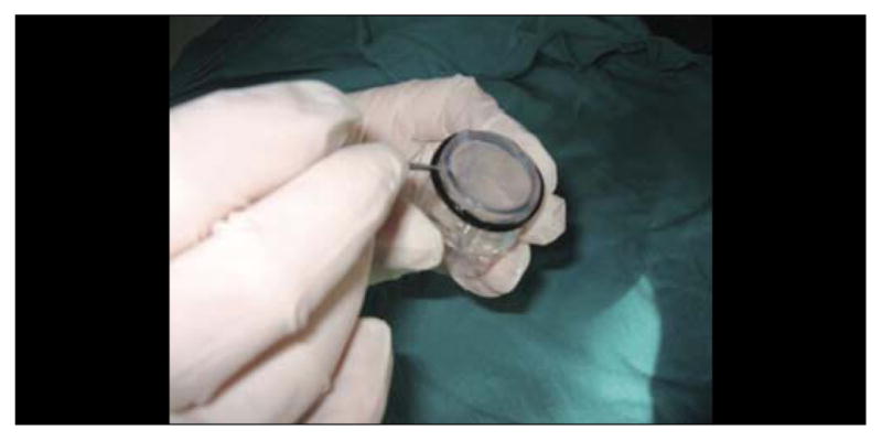
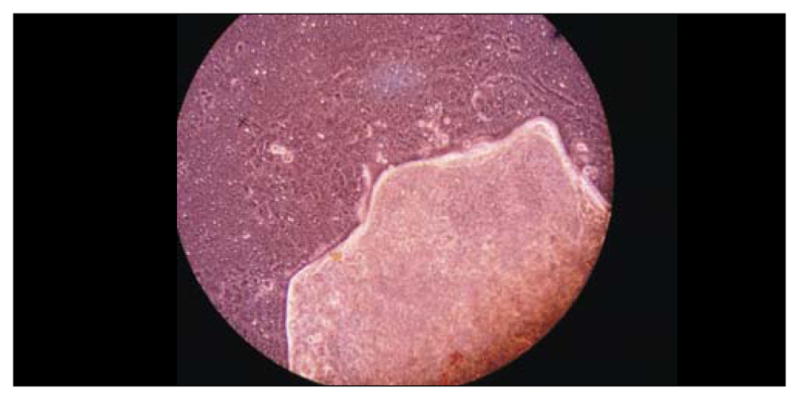
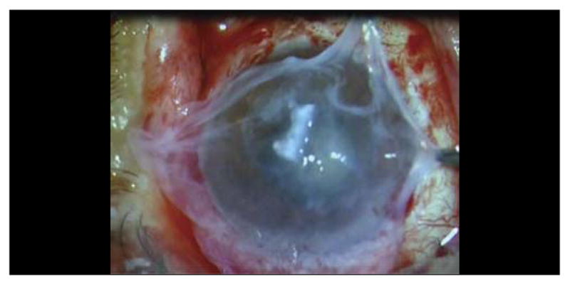
One-year follow-up results of five unilateral LSCD patients who had undergone CLEC transplantation were evaluated. Parameters for this evaluation were: fluorescein staining of ocular surface, corneal vascularization and status of epithelium with slit lamp, and visual acuity. 1.5-mm limbal biopsy was performed from the superior limbus of the healthy eyes, broke into two equal pieces, expanded on human amniotic membrane (hAM) and inserts for 14 days until getting 20 mm in size. CLECs on hAMs were used directly, and cells on inserts were used after detachment procedure. The symblepharon and pannus tissues were removed, superficial keratectomy was performed. CLEC on hAMs were transplanted with the epithelial side up onto the bare corneal stroma, sutured to the conjunctiva with 10-0 nylon sutures. Free CLEC layer from insert was placed on hAM as a second layer, additional hAM was used as a protective layer all over other tissues.

RESULTS

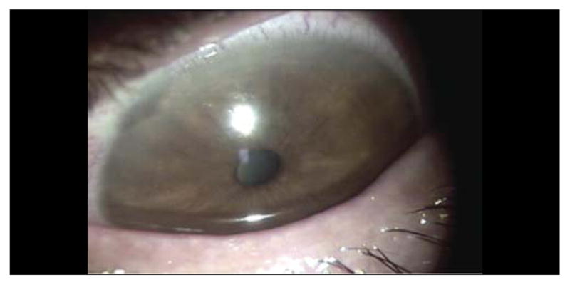
Median age was 44.4 years (14-71). The etiology was chemical burn in all patients. Median duration of symptoms was 10 years (2-18), median follow-up period was 12.6 (12-12.5) months. Preoperative best corrected visual acuities (BCVA) were light perception in three patients, counting fingers at 50 cm in one patient and 3/10 in one patient. Visions were improved in all patients. Postoperative BCVA 12 months after the surgery were between counting fingers at 3 meters to 6/10. There was a temporary hemorrhage between the two layers of hAMs in one patient at the early postoperative period. Peripheral corneal vascularization has occurred in three patients, in patient corneal vascularization has reached to the paracentral area.

DISCUSSION

CLEC transplantation is an efficient treatment option for unilateral LSCD in mid-long term.

摘要

目的

评估培养的角膜缘上皮细胞(CLEC)移植治疗单侧角膜缘干细胞缺乏症(LSCD)的1年随访结果。

材料与方法

对5例接受CLEC移植的单侧LSCD患者进行1年随访评估。评估参数包括:眼表荧光素染色、角膜血管化情况、裂隙灯检查下的上皮状态以及视力。从健康眼的上方角膜缘取1.5mm的角膜缘活检组织,分成两块相等的组织块,在人羊膜(hAM)上进行扩增并培养14天,直至其大小达到20mm。直接使用hAM上的CLEC,将培养皿上的细胞在分离后使用。切除睑球粘连和血管翳组织,进行浅层角膜切除术。将hAM上的CLEC上皮面朝上移植到裸露的角膜基质上,用10-0尼龙缝线缝合到结膜上。将培养皿上的游离CLEC层作为第二层放置在hAM上,额外的hAM用作覆盖其他组织的保护层。

结果

中位年龄为44.4岁(14-71岁)。所有患者的病因均为化学伤。症状的中位持续时间为10年(2-18年),中位随访期为12.6(12-12.5)个月。术前最佳矫正视力(BCVA):3例患者为光感,1例患者为50cm处数指,1例患者为3/10。所有患者的视力均有改善。术后12个月的BCVA在3米处数指至6/10之间。1例患者在术后早期两层hAM之间出现暂时性出血。3例患者发生周边角膜血管化,其中1例患者的角膜血管化已延伸至旁中央区。

讨论

CLEC移植是治疗单侧LSCD的中长期有效治疗选择。

https://cdn.ncbi.nlm.nih.gov/pmc/blobs/c26d/5576176/b3cf697413b7/nihms889390f6.jpg
https://cdn.ncbi.nlm.nih.gov/pmc/blobs/c26d/5576176/a9903d0da1e0/nihms889390f1.jpg
https://cdn.ncbi.nlm.nih.gov/pmc/blobs/c26d/5576176/54c27a392a94/nihms889390f2.jpg
https://cdn.ncbi.nlm.nih.gov/pmc/blobs/c26d/5576176/6a82ad8cba0c/nihms889390f3.jpg
https://cdn.ncbi.nlm.nih.gov/pmc/blobs/c26d/5576176/8cf4797f540c/nihms889390f4.jpg
https://cdn.ncbi.nlm.nih.gov/pmc/blobs/c26d/5576176/254bbfb1107e/nihms889390f5.jpg
https://cdn.ncbi.nlm.nih.gov/pmc/blobs/c26d/5576176/b3cf697413b7/nihms889390f6.jpg

相似文献

1
[Ocular Surface Reconstruction with Cultivated Limbal Epithelial Cells in Limbal Stem Cell Deficiency: One-year Follow-up Results].[培养的角膜缘上皮细胞用于角膜缘干细胞缺乏症的眼表重建:一年随访结果]
Turk Oftalmol Derg. 2012 May;42(3):172-176. doi: 10.4274/tjo.42.66375.
2
[Amniotic membrane graft in ocular surface disease. Prospective study with 31 cases].[羊膜移植治疗眼表疾病。31例前瞻性研究]
J Fr Ophtalmol. 2001 Oct;24(8):798-812.
3
Amniotic membrane transplantation with conjunctival limbal autograft for total limbal stem cell deficiency.羊膜移植联合自体结膜角膜缘移植治疗全角膜缘干细胞缺乏症
Ophthalmology. 2003 Aug;110(8):1585-92. doi: 10.1016/S0161-6420(03)00503-7.
4
Reconstruction of damaged corneas by transplantation of autologous limbal epithelial cells.通过自体角膜缘上皮细胞移植重建受损角膜。
N Engl J Med. 2000 Jul 13;343(2):86-93. doi: 10.1056/NEJM200007133430202.
5
Use of autologous cultured limbal and conjunctival epithelium in a patient with severe bilateral ocular surface disease induced by acid injury: a case report of unique application.自体培养角膜缘和结膜上皮在酸烧伤所致严重双侧眼表疾病患者中的应用:独特应用的病例报告
Cornea. 2003 Jul;22(5):478-81. doi: 10.1097/00003226-200307000-00016.
6
Limbal stem cell and oral mucosal epithelial transplantation from ex vivo cultivation in LSCD-induced rabbits: histology and immunologic study of the transplant epithelial sheet.体外培养的角膜缘干细胞和口腔黏膜上皮移植于兔角膜缘干细胞缺乏诱导模型:移植上皮片的组织学和免疫学研究
Int Ophthalmol. 2017 Dec;37(6):1289-1298. doi: 10.1007/s10792-016-0402-5. Epub 2016 Dec 2.
7
Midterm results of cultivated autologous and allogeneic limbal epithelial transplantation in limbal stem cell deficiency.自体和异体培养角膜缘上皮移植治疗角膜缘干细胞缺乏症的中期结果
Dev Ophthalmol. 2010;45:57-70. doi: 10.1159/000315020. Epub 2010 May 18.
8
Application of autologous cultivated corneal epithelium for corneal limbal stem cell insufficiency--short-term results.自体培养角膜上皮在角膜缘干细胞缺乏症中的应用——短期结果
Klin Oczna. 2011;113(10-12):346-51.
9
Characterization of the corneal surface in limbal stem cell deficiency and after transplantation of cultivated limbal epithelium.角膜缘干细胞缺乏症及培养角膜缘上皮移植后的角膜表面特征
Ophthalmology. 2009 Jun;116(6):1048-56. doi: 10.1016/j.ophtha.2009.01.005. Epub 2009 Apr 25.
10
[Transplantation of human limbal cells cultivated on amniotic membrane for reconstruction of rat corneal epithelium after alkali burn].[羊膜上培养的人角膜缘细胞移植用于碱烧伤大鼠角膜上皮重建]
Zhonghua Yan Ke Za Zhi. 2007 Feb;43(2):134-41.

引用本文的文献

1
Limbal Stem Cell Deficiency and Treatment with Stem Cell Transplantation.角膜缘干细胞缺乏症与干细胞移植治疗
Turk J Ophthalmol. 2017 Oct;47(5):285-291. doi: 10.4274/tjo.72593. Epub 2017 Oct 27.
2
Clinical Spectrum and Treatment Approaches in Corneal Burns.角膜烧伤的临床谱及治疗方法
Turk J Ophthalmol. 2015 Oct;45(5):182-187. doi: 10.4274/tjo.99267. Epub 2015 Oct 5.

本文引用的文献

1
Ocular surface reconstruction with cultivated limbal epithelium in a patient with unilateral stem cell deficiency caused by Epidermolysis bullosa dystrophica hallopeau-Siemens.用培养的角膜缘上皮细胞对因营养不良型大疱性表皮松解症导致的单侧干细胞缺乏症患者进行眼表面重建。
Cornea. 2010 Apr;29(4):462-4. doi: 10.1097/ICO.0b013e3181b442ea.
2
Successful clinical implementation of corneal epithelial stem cell therapy for treatment of unilateral limbal stem cell deficiency.成功地将角膜上皮干细胞治疗应用于单侧角膜缘干细胞缺陷的临床治疗。
Stem Cells. 2010 Mar 31;28(3):597-610. doi: 10.1002/stem.276.
3
Cultivated limbal stem cell transplantation for ocular surface reconstruction.
培养的角膜缘干细胞移植用于眼表重建。
Clin Ophthalmol. 2008 Sep;2(3):489-502.
4
Limbal stem cell transplantation: new progresses and challenges.角膜缘干细胞移植:新进展与挑战。
Eye (Lond). 2009 Oct;23(10):1946-53. doi: 10.1038/eye.2008.379. Epub 2008 Dec 19.
5
Clinical outcome of autologous cultivated limbal epithelium transplantation.自体培养角膜缘上皮移植的临床结果
Indian J Ophthalmol. 2006 Mar;54(1):29-34. doi: 10.4103/0301-4738.21611.
6
Early results of penetrating keratoplasty after cultivated limbal epithelium transplantation.培养的角膜缘上皮移植术后穿透性角膜移植术的早期结果
Arch Ophthalmol. 2005 Mar;123(3):334-40. doi: 10.1001/archopht.123.3.334.
7
Corneal stromal changes following reconstruction by ex vivo expanded limbal epithelial cells in rabbits with total limbal stem cell deficiency.在完全角膜缘干细胞缺乏的兔中,经体外扩增角膜缘上皮细胞重建后角膜基质的变化。
Br J Ophthalmol. 2003 Dec;87(12):1509-14. doi: 10.1136/bjo.87.12.1509.
8
Successful regrafting of cultivated corneal epithelium using amniotic membrane as a carrier in severe ocular surface disease.在严重眼表疾病中,以羊膜为载体成功进行培养角膜上皮的再移植。
Cornea. 2003 Jan;22(1):70-1. doi: 10.1097/00003226-200301000-00017.
9
Phenotypic study of a case with successful transplantation of ex vivo expanded human limbal epithelium for unilateral total limbal stem cell deficiency.1例单侧完全性角膜缘干细胞缺乏患者成功移植体外扩增人角膜缘上皮的表型研究
Ophthalmology. 2002 Aug;109(8):1547-52. doi: 10.1016/s0161-6420(02)01105-3.
10
Human limbal progenitor cells expanded on intact amniotic membrane ex vivo.人角膜缘祖细胞在完整羊膜上进行体外扩增。
Arch Ophthalmol. 2002 Jun;120(6):783-90. doi: 10.1001/archopht.120.6.783.