• 文献检索
  • 文档翻译
  • 深度研究
  • 学术资讯
  • Suppr Zotero 插件Zotero 插件
  • 邀请有礼
  • 套餐&价格
  • 历史记录
应用&插件
Suppr Zotero 插件Zotero 插件浏览器插件Mac 客户端Windows 客户端微信小程序
定价
高级版会员购买积分包购买API积分包
服务
文献检索文档翻译深度研究API 文档MCP 服务
关于我们
关于 Suppr公司介绍联系我们用户协议隐私条款
关注我们

Suppr 超能文献

核心技术专利:CN118964589B侵权必究
粤ICP备2023148730 号-1Suppr @ 2026

文献检索

告别复杂PubMed语法,用中文像聊天一样搜索,搜遍4000万医学文献。AI智能推荐,让科研检索更轻松。

立即免费搜索

文件翻译

保留排版,准确专业,支持PDF/Word/PPT等文件格式,支持 12+语言互译。

免费翻译文档

深度研究

AI帮你快速写综述,25分钟生成高质量综述,智能提取关键信息,辅助科研写作。

立即免费体验

可用于鉴别小的三阴性乳腺癌和纤维腺瘤的超声特征。

Sonographic features that can be used to differentiate between small triple-negative breast cancer and fibroadenoma.

作者信息

Yoon Ga Young, Cha Joo Hee, Kim Hak Hee, Shin Hee Jung, Chae Eun Young, Choi Woo Jung

机构信息

Department of Radiology and Research Institute of Radiology, Asan Medical Center, University of Ulsan College of Medicine, Seoul, Korea.

出版信息

Ultrasonography. 2018 Apr;37(2):149-156. doi: 10.14366/usg.17036. Epub 2017 Aug 4.

DOI:10.14366/usg.17036
PMID:28870060
原文链接:https://pmc.ncbi.nlm.nih.gov/articles/PMC5885477/
Abstract

PURPOSE

Our study investigated whether any sonographic findings could be useful for differentiating between small triple-negative breast cancer (TNBC) and fibroadenoma.

METHODS

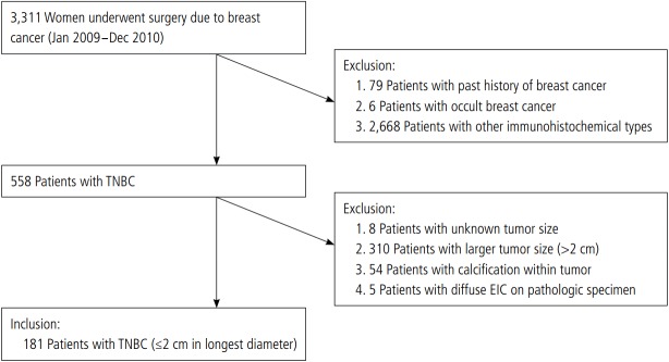
This retrospective study was approved by our Institutional Review Board, which waived the requirement for patient consent. From January 2009 to December 2010, the sonographic features of 181 pathologically proven TNBC tumors and 172 fibroadenomas measuring less than or equal to 2 cm in the longest dimension were reviewed and analyzed according to the fifth edition of the Breast Imaging Reporting and Data System (BI-RADS) lexicon. Mean tumor roundness was also measured using in-house software.

RESULTS

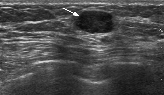
The median longest lesion dimension was 16 mm (range, 13 to 18 mm) in TNBCs and 13 mm (range, 10 to 16 mm) in fibroadenomas. In comparison to fibroadenomas, small TNBC tumors presented with a higher incidence of irregular shapes (66.9%), noncircumscribed margins (91.7%), hypoechoic echotexture (59.1%), posterior acoustic enhancement (65.2%), and associated features (24.4%). Most TNBCs were classified as BI-RADS category 4 (65.2%) or 5 (28.2%). The mean tumor roundness of small TNBCs was greater than that of fibroadenomas (60%±12% vs. 53%±13%). Multivariate analysis showed that older patient age, irregular shape, nonparallel orientation, posterior acoustic enhancement, associated features, a BI-RADS final assessment category of 4 or 5, and greater tumor roundness were significant independent factors indicative of TNBCs.

CONCLUSION

TNBC tumors tend to demonstrate more suspicious sonographic features and greater tumor roundness than fibroadenomas. These features may have the potential to help differentiate between small TNBCs and fibroadenomas.

摘要

目的

我们的研究调查了是否有任何超声检查结果可用于鉴别小的三阴性乳腺癌(TNBC)和纤维腺瘤。

方法

这项回顾性研究经我们机构审查委员会批准,该委员会免除了患者知情同意的要求。2009年1月至2010年12月,根据《乳腺影像报告和数据系统(BI-RADS)》第五版词汇表,对181例经病理证实的TNBC肿瘤和172例最大径小于或等于2 cm的纤维腺瘤的超声特征进行了回顾和分析。还使用内部软件测量了肿瘤的平均圆度。

结果

TNBC的病变最长径中位数为16 mm(范围13至18 mm),纤维腺瘤为13 mm(范围10至16 mm)。与纤维腺瘤相比,小的TNBC肿瘤具有更高的不规则形状发生率(66.9%)、非边缘清晰的边界(91.7%)、低回声质地(59.1%)、后方回声增强(65.2%)以及相关特征(24.4%)。大多数TNBC被分类为BI-RADS 4类(65.2%)或5类(28.2%)。小TNBC的平均肿瘤圆度大于纤维腺瘤(60%±12%对53%±13%)。多变量分析显示,患者年龄较大、形状不规则、非平行方向、后方回声增强、相关特征、BI-RADS最终评估类别为4或5以及更大的肿瘤圆度是提示TNBC的显著独立因素。

结论

与纤维腺瘤相比,TNBC肿瘤往往表现出更可疑的超声特征和更大的肿瘤圆度。这些特征可能有助于鉴别小的TNBC和纤维腺瘤。

https://cdn.ncbi.nlm.nih.gov/pmc/blobs/d0c1/5885477/f246a4bd2e77/usg-17036-f2.jpg
https://cdn.ncbi.nlm.nih.gov/pmc/blobs/d0c1/5885477/eecb21bc2afd/usg-17036-f1.jpg
https://cdn.ncbi.nlm.nih.gov/pmc/blobs/d0c1/5885477/f246a4bd2e77/usg-17036-f2.jpg
https://cdn.ncbi.nlm.nih.gov/pmc/blobs/d0c1/5885477/eecb21bc2afd/usg-17036-f1.jpg
https://cdn.ncbi.nlm.nih.gov/pmc/blobs/d0c1/5885477/f246a4bd2e77/usg-17036-f2.jpg

相似文献

1
Sonographic features that can be used to differentiate between small triple-negative breast cancer and fibroadenoma.可用于鉴别小的三阴性乳腺癌和纤维腺瘤的超声特征。
Ultrasonography. 2018 Apr;37(2):149-156. doi: 10.14366/usg.17036. Epub 2017 Aug 4.
2
Sonographic appearances of juvenile fibroadenoma of the breast.乳腺幼年型纤维腺瘤的超声表现。
J Ultrasound Med. 2014 Nov;33(11):1879-84. doi: 10.7863/ultra.33.11.1879.
3
Gray-scale and color Doppler sonographic features of complex fibroadenomas of the breast.乳腺复杂性纤维腺瘤的灰阶及彩色多普勒超声特征
J Clin Ultrasound. 2015 Nov-Dec;43(9):556-62. doi: 10.1002/jcu.22278. Epub 2015 Jul 21.
4
Breast fibroadenomas in the pediatric population: common and uncommon sonographic findings.儿科人群中的乳腺纤维腺瘤:常见和不常见的超声表现。
Pediatr Radiol. 2010 Oct;40(10):1681-9. doi: 10.1007/s00247-010-1678-7. Epub 2010 May 7.
5
Simple and complex fibroadenomas: are there any distinguishing sonographic features?单纯性和复杂性纤维腺瘤:是否存在任何有鉴别意义的超声特征?
J Ultrasound Med. 2014 Mar;33(3):415-9. doi: 10.7863/ultra.33.3.415.
6
Prediction for pathological and immunohistochemical characteristics of triple-negative invasive breast carcinomas: the performance comparison between quantitative and qualitative sonographic feature analysis.三阴性浸润性乳腺癌病理及免疫组织化学特征预测:定量与定性超声特征分析的性能比较。
Eur Radiol. 2022 Mar;32(3):1590-1600. doi: 10.1007/s00330-021-08224-x. Epub 2021 Sep 14.
7
Texture Analysis of Ultrasound Images to Differentiate Simple Fibroadenomas From Complex Fibroadenomas and Benign Phyllodes Tumors.超声图像纹理分析用于鉴别单纯性纤维腺瘤与复杂性纤维腺瘤及良性叶状肿瘤
J Ultrasound Med. 2020 Oct;39(10):1993-2003. doi: 10.1002/jum.15304. Epub 2020 Apr 24.
8
Nonpalpable BI-RADS 4 breast lesions: sonographic findings and pathology correlation.不可触及的乳腺影像报告和数据系统(BI-RADS)4类乳腺病变:超声表现与病理相关性
Diagn Interv Radiol. 2015 May-Jun;21(3):189-94. doi: 10.5152/dir.2014.14103.
9
The potential role of breast MRI in evaluation of triple-negative breast cancer and fibroadenoma of less than 3 cm.乳腺磁共振成像在评估三阴性乳腺癌及小于3厘米的纤维腺瘤中的潜在作用
Transl Cancer Res. 2024 Aug 31;13(8):4042-4051. doi: 10.21037/tcr-24-498. Epub 2024 Aug 27.
10
Ultrasonographic features of triple-negative breast cancer: a comparison with other breast cancer subtypes.三阴性乳腺癌的超声特征:与其他乳腺癌亚型的比较。
Asian Pac J Cancer Prev. 2015;16(8):3229-32. doi: 10.7314/apjcp.2015.16.8.3229.

引用本文的文献

1
Identification of differentiating sonographic features between fibroadenomas and malignant tumors of the breast mimicking fibroadenomas: 10-year experience in 421 histologically verified cases.鉴别纤维腺瘤与酷似纤维腺瘤的乳腺恶性肿瘤之间的超声鉴别特征:421例经组织学证实病例的10年经验
Ultraschall Med. 2025 Jun;46(3):291-298. doi: 10.1055/a-2474-6617. Epub 2025 Feb 4.
2
The value of CT shape quantification in predicting pathological classification of lung adenocarcinoma.CT 形态学定量在预测肺腺癌病理分类中的价值。
BMC Cancer. 2024 Jan 4;24(1):35. doi: 10.1186/s12885-023-11802-5.
3
Prevalence of breast fibroadenoma in healthy physical examination population in Guangdong province of China: a cross-sectional study.

本文引用的文献

1
Differences in Multi-Modal Ultrasound Imaging between Triple Negative and Non-Triple Negative Breast Cancer.三阴性乳腺癌与非三阴性乳腺癌的多模态超声成像差异
Ultrasound Med Biol. 2016 Apr;42(4):882-90. doi: 10.1016/j.ultrasmedbio.2015.12.003. Epub 2016 Jan 16.
2
Ultrasonographic findings of triple-negative breast cancer.三阴性乳腺癌的超声检查结果。
Int J Clin Exp Med. 2015 Jun 15;8(6):10040-3. eCollection 2015.
3
Computer-aided diagnosis for distinguishing between triple-negative breast cancer and fibroadenomas based on ultrasound texture features.
中国广东省健康体检人群中乳腺纤维腺瘤的患病率:一项横断面研究。
BMJ Open. 2022 Jun 27;12(6):e057080. doi: 10.1136/bmjopen-2021-057080.
4
Value of Histogram of Gray-Scale Ultrasound Image in Differential Diagnosis of Small Triple Negative Breast Invasive Ductal Carcinoma and Fibroadenoma.灰度超声图像直方图在三阴性乳腺浸润性导管癌与纤维瘤鉴别诊断中的价值
Cancer Manag Res. 2022 Apr 21;14:1515-1524. doi: 10.2147/CMAR.S359986. eCollection 2022.
5
Ultrasound-based radiomics nomogram for differentiation of triple-negative breast cancer from fibroadenoma.基于超声的放射组学列线图用于鉴别三阴性乳腺癌与纤维腺瘤。
Br J Radiol. 2022 May 1;95(1133):20210598. doi: 10.1259/bjr.20210598. Epub 2022 Feb 9.
6
Characterization of soft tissue tumours with ultrasound, shear wave elastography and MRI.超声、剪切波弹性成像及 MRI 对软组织肿瘤的特征描述。
Skeletal Radiol. 2020 Jun;49(6):869-881. doi: 10.1007/s00256-019-03363-1. Epub 2020 Jan 2.
7
Radiomics of US texture features in differential diagnosis between triple-negative breast cancer and fibroadenoma.超声纹理特征在三阴性乳腺癌与纤维腺瘤鉴别诊断中的放射组学研究。
Sci Rep. 2018 Sep 10;8(1):13546. doi: 10.1038/s41598-018-31906-4.
基于超声纹理特征的三阴性乳腺癌与纤维腺瘤的计算机辅助诊断
Med Phys. 2015 Jun;42(6):3024-35. doi: 10.1118/1.4921123.
4
Outcomes of solid palpable masses assessed as BI-RADS 3 or 4A: a retrospective review.评估为BI-RADS 3或4A的可触及实性肿块的结局:一项回顾性研究。
Breast Cancer Res Treat. 2014 Sep;147(2):311-6. doi: 10.1007/s10549-014-3109-1. Epub 2014 Aug 24.
5
Tumour volume doubling time of molecular breast cancer subtypes assessed by serial breast ultrasound.通过连续乳腺超声评估的分子乳腺癌亚型的肿瘤体积倍增时间
Eur Radiol. 2014 Sep;24(9):2227-35. doi: 10.1007/s00330-014-3256-0. Epub 2014 Jun 4.
6
Quantitative MRI morphology of invasive breast cancer: correlation with immunohistochemical biomarkers and subtypes.浸润性乳腺癌的定量MRI形态学:与免疫组化生物标志物及亚型的相关性
Acta Radiol. 2015 Mar;56(3):269-75. doi: 10.1177/0284185114524197. Epub 2014 Feb 20.
7
Breast cancer detected with screening US: reasons for nondetection at mammography.用筛查超声检测出的乳腺癌:乳腺 X 线摄影漏诊的原因。
Radiology. 2014 Feb;270(2):369-77. doi: 10.1148/radiol.13130724. Epub 2013 Nov 6.
8
Imaging of triple-negative breast cancer.三阴性乳腺癌的影像学表现。
Ann Oncol. 2012 Aug;23 Suppl 6:vi23-9. doi: 10.1093/annonc/mds191.
9
Sonographic features of histopathologically benign solid breast lesions that have been classified as BI-RADS 4 on sonography.超声检查分类为BI-RADS 4类的组织病理学良性实性乳腺病变的超声特征。
J Clin Ultrasound. 2012 Jun;40(5):261-5. doi: 10.1002/jcu.21923. Epub 2012 Apr 17.
10
Speckle reduction imaging of breast ultrasound does not improve the diagnostic performance of morphology-based CAD System.乳腺超声斑点减少成像并不能提高基于形态学的计算机辅助检测系统的诊断性能。
J Clin Ultrasound. 2012 Jan;40(1):1-6. doi: 10.1002/jcu.20897. Epub 2011 Nov 15.