Department of Radiology, Severance Hospital, Research Institute of Radiological Science and Center for Clinical Image Data Science, Yonsei University College of Medicine, Seoul, Korea.
Department of Computational Science and Engineering, Yonsei University, Seoul, Korea.
Sci Rep. 2018 Sep 10;8(1):13546. doi: 10.1038/s41598-018-31906-4.
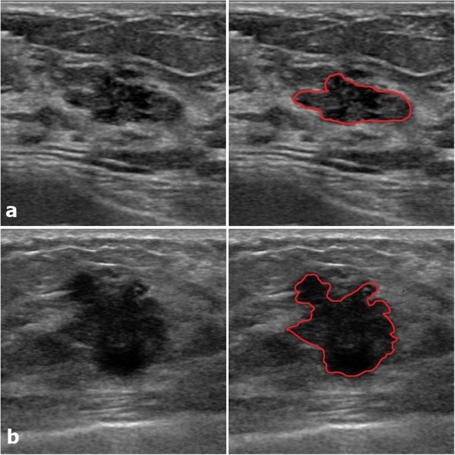
Triple-negative breast cancer (TNBC) is sometimes mistaken for fibroadenoma due to its tendency to show benign morphology on breast ultrasound (US) albeit its aggressive nature. This study aims to develop a radiomics score based on US texture analysis for differential diagnosis between TNBC and fibroadenoma, and to evaluate its diagnostic performance compared with pathologic results. We retrospectively included 715 pathology-proven fibroadenomas and 186 pathology-proven TNBCs which were examined by three different US machines. We developed the radiomics score by using penalized logistic regression with a least absolute shrinkage and selection operator (LASSO) analysis from 730 extracted features consisting of 14 intensity-based features, 132 textural features and 584 wavelet-based features. The constructed radiomics score showed significant difference between fibroadenoma and TNBC for all three US machines (p < 0.001). Although the radiomics score showed dependency on the type of US machine, we developed more elaborate radiomics score for a subgroup in which US examinations were performed with iU22. This subsequent radiomics score also showed good diagnostic performance, even for BI-RADS category 3 or 4a lesions (AUC 0.782) which were presumed as probably benign or low suspicious of malignancy by radiologists. It was expected to assist radiologist's diagnosis and reduce the number of invasive biopsies, although US standardization should be overcome before clinical application.
三阴性乳腺癌(TNBC)在乳腺超声(US)上表现出良性形态,容易被误诊为纤维腺瘤,尽管其具有侵袭性。本研究旨在基于 US 纹理分析开发一种用于鉴别 TNBC 和纤维腺瘤的放射组学评分,并评估其与病理结果相比的诊断性能。我们回顾性纳入了 715 例经病理证实的纤维腺瘤和 186 例经病理证实的 TNBC,这些患者均由 3 台不同的 US 机器进行检查。我们通过使用惩罚逻辑回归和最小绝对收缩和选择算子(LASSO)分析从 730 个提取特征中开发了放射组学评分,这些特征包括 14 个基于强度的特征、132 个纹理特征和 584 个基于小波的特征。该构建的放射组学评分在所有 3 台 US 机器上均显示出纤维腺瘤和 TNBC 之间的显著差异(p<0.001)。尽管放射组学评分显示对 US 机器类型存在依赖性,但我们为使用 iU22 进行 US 检查的亚组开发了更精细的放射组学评分。该后续放射组学评分也表现出良好的诊断性能,甚至对 BI-RADS 类别为 3 或 4a 的病变(AUC 0.782)也有很好的诊断性能,这些病变被放射科医生认为可能是良性或低度恶性可疑。虽然在临床应用之前应该克服 US 标准化问题,但该方法有望协助放射科医生的诊断并减少有创活检的数量。