• 文献检索
  • 文档翻译
  • 深度研究
  • 学术资讯
  • Suppr Zotero 插件Zotero 插件
  • 邀请有礼
  • 套餐&价格
  • 历史记录
应用&插件
Suppr Zotero 插件Zotero 插件浏览器插件Mac 客户端Windows 客户端微信小程序
定价
高级版会员购买积分包购买API积分包
服务
文献检索文档翻译深度研究API 文档MCP 服务
关于我们
关于 Suppr公司介绍联系我们用户协议隐私条款
关注我们

Suppr 超能文献

核心技术专利:CN118964589B侵权必究
粤ICP备2023148730 号-1Suppr @ 2026

文献检索

告别复杂PubMed语法,用中文像聊天一样搜索,搜遍4000万医学文献。AI智能推荐,让科研检索更轻松。

立即免费搜索

文件翻译

保留排版,准确专业,支持PDF/Word/PPT等文件格式,支持 12+语言互译。

免费翻译文档

深度研究

AI帮你快速写综述,25分钟生成高质量综述,智能提取关键信息,辅助科研写作。

立即免费体验

利用专家分割标签和放射组学特征推进癌症基因组图谱胶质细胞瘤 MRI 数据集。

Advancing The Cancer Genome Atlas glioma MRI collections with expert segmentation labels and radiomic features.

机构信息

Center for Biomedical Image Computing and Analytics (CBICA), Perelman School of Medicine, University of Pennsylvania, Richards Medical Research Laboratories, Floor 7, 3700 Hamilton Walk, Philadelphia, Pennsylvania 19104, USA.

Department of Radiology, Perelman School of Medicine, University of Pennsylvania, Richards Medical Research Laboratories, Floor 7, 3700 Hamilton Walk, Philadelphia, Pennsylvania 19104, USA.

出版信息

Sci Data. 2017 Sep 5;4:170117. doi: 10.1038/sdata.2017.117.

DOI:10.1038/sdata.2017.117
PMID:28872634
原文链接:https://pmc.ncbi.nlm.nih.gov/articles/PMC5685212/
Abstract

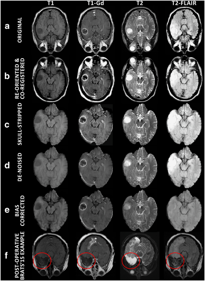
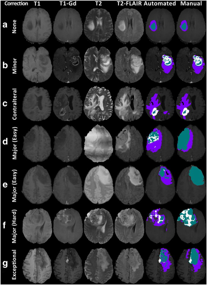
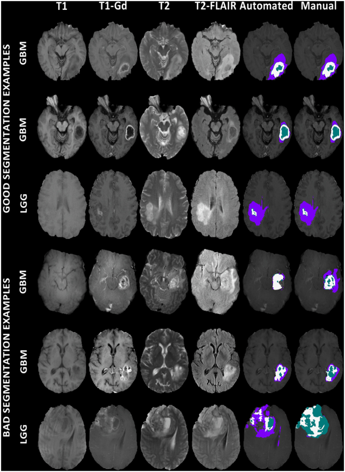
Gliomas belong to a group of central nervous system tumors, and consist of various sub-regions. Gold standard labeling of these sub-regions in radiographic imaging is essential for both clinical and computational studies, including radiomic and radiogenomic analyses. Towards this end, we release segmentation labels and radiomic features for all pre-operative multimodal magnetic resonance imaging (MRI) (n=243) of the multi-institutional glioma collections of The Cancer Genome Atlas (TCGA), publicly available in The Cancer Imaging Archive (TCIA). Pre-operative scans were identified in both glioblastoma (TCGA-GBM, n=135) and low-grade-glioma (TCGA-LGG, n=108) collections via radiological assessment. The glioma sub-region labels were produced by an automated state-of-the-art method and manually revised by an expert board-certified neuroradiologist. An extensive panel of radiomic features was extracted based on the manually-revised labels. This set of labels and features should enable i) direct utilization of the TCGA/TCIA glioma collections towards repeatable, reproducible and comparative quantitative studies leading to new predictive, prognostic, and diagnostic assessments, as well as ii) performance evaluation of computer-aided segmentation methods, and comparison to our state-of-the-art method.

摘要

神经胶质瘤属于中枢神经系统肿瘤的一种,由各种不同的子区域组成。在放射影像学中对这些子区域进行金标准标记对于临床和计算研究都至关重要,包括放射组学和放射基因组学分析。为此,我们发布了来自癌症基因组图谱(TCGA)多机构神经胶质瘤数据集的所有术前多模态磁共振成像(MRI)(n=243)的分割标签和放射组学特征,这些数据可在癌症成像档案库(TCIA)中公开获取。术前扫描通过放射学评估在胶质母细胞瘤(TCGA-GBM,n=135)和低级别神经胶质瘤(TCGA-LGG,n=108)两个集合中被识别出来。胶质瘤子区域标签由自动化的最先进方法生成,并由经过专业认证的神经放射科医生进行手动修订。根据手动修订的标签提取了大量的放射组学特征。这组标签和特征应能 i)直接利用 TCGA/TCIA 神经胶质瘤数据集进行可重复、可重现和可比的定量研究,从而实现新的预测、预后和诊断评估,以及 ii)评估计算机辅助分割方法的性能,并与我们的最先进方法进行比较。

https://cdn.ncbi.nlm.nih.gov/pmc/blobs/5728/5685212/140ebfc034c9/sdata2017117-f3.jpg
https://cdn.ncbi.nlm.nih.gov/pmc/blobs/5728/5685212/9ac77033dd85/sdata2017117-f1.jpg
https://cdn.ncbi.nlm.nih.gov/pmc/blobs/5728/5685212/88782a88389c/sdata2017117-f2.jpg
https://cdn.ncbi.nlm.nih.gov/pmc/blobs/5728/5685212/140ebfc034c9/sdata2017117-f3.jpg
https://cdn.ncbi.nlm.nih.gov/pmc/blobs/5728/5685212/9ac77033dd85/sdata2017117-f1.jpg
https://cdn.ncbi.nlm.nih.gov/pmc/blobs/5728/5685212/88782a88389c/sdata2017117-f2.jpg
https://cdn.ncbi.nlm.nih.gov/pmc/blobs/5728/5685212/140ebfc034c9/sdata2017117-f3.jpg

相似文献

1
Advancing The Cancer Genome Atlas glioma MRI collections with expert segmentation labels and radiomic features.利用专家分割标签和放射组学特征推进癌症基因组图谱胶质细胞瘤 MRI 数据集。
Sci Data. 2017 Sep 5;4:170117. doi: 10.1038/sdata.2017.117.
2
Machine learning and radiomic phenotyping of lower grade gliomas: improving survival prediction.机器学习和低级别胶质瘤的放射组学表型:改善生存预测。
Eur Radiol. 2020 Jul;30(7):3834-3842. doi: 10.1007/s00330-020-06737-5. Epub 2020 Mar 11.
3
Genotype prediction of ATRX mutation in lower-grade gliomas using an MRI radiomics signature.使用 MRI 放射组学特征预测低级别胶质瘤中的 ATRX 突变。
Eur Radiol. 2018 Jul;28(7):2960-2968. doi: 10.1007/s00330-017-5267-0. Epub 2018 Feb 5.
4
Magnetic resonance imaging-based radiomic features for extrapolating infiltration levels of immune cells in lower-grade gliomas.基于磁共振成像的放射组学特征可推断低级别胶质瘤中免疫细胞浸润程度。
Strahlenther Onkol. 2020 Oct;196(10):913-921. doi: 10.1007/s00066-020-01584-1. Epub 2020 Feb 5.
5
Reproducibility analysis of multi-institutional paired expert annotations and radiomic features of the Ivy Glioblastoma Atlas Project (Ivy GAP) dataset.多机构配对专家注释和 Ivy 脑胶质瘤图谱项目 (Ivy GAP) 数据集放射组学特征的可重复性分析。
Med Phys. 2020 Dec;47(12):6039-6052. doi: 10.1002/mp.14556. Epub 2020 Dec 4.
6
Comprehensive quantitative radiogenomic evaluation reveals novel radiomic subtypes with distinct immune pattern in glioma.全面定量放射基因组学评估揭示了胶质瘤中具有不同免疫模式的新型放射组学分型。
Comput Biol Med. 2024 Jul;177:108636. doi: 10.1016/j.compbiomed.2024.108636. Epub 2024 May 20.
7
Comparison of unsupervised classification methods for brain tumor segmentation using multi-parametric MRI.使用多参数磁共振成像的脑肿瘤分割无监督分类方法比较
Neuroimage Clin. 2016 Sep 30;12:753-764. doi: 10.1016/j.nicl.2016.09.021. eCollection 2016.
8
Brain extraction on MRI scans in presence of diffuse glioma: Multi-institutional performance evaluation of deep learning methods and robust modality-agnostic training.MRI 扫描中存在弥漫性胶质瘤时的脑提取:深度学习方法的多机构性能评估和稳健的模态不可知训练。
Neuroimage. 2020 Oct 15;220:117081. doi: 10.1016/j.neuroimage.2020.117081. Epub 2020 Jun 27.
9
A Coclinical Radiogenomic Validation Study: Conserved Magnetic Resonance Radiomic Appearance of Periostin-Expressing Glioblastoma in Patients and Xenograft Models.一项临床放射组学验证研究:成纤维细胞激活蛋白表达胶质母细胞瘤患者和异种移植模型磁共振放射组学表现的保守性。
Clin Cancer Res. 2018 Dec 15;24(24):6288-6299. doi: 10.1158/1078-0432.CCR-17-3420. Epub 2018 Jul 27.
10
Radiogenomic-Based Survival Risk Stratification of Tumor Habitat on Gd-T1w MRI Is Associated with Biological Processes in Glioblastoma.基于影像组学的钆增强 T1wMRI 肿瘤微环境生存风险分层与胶质母细胞瘤的生物学过程相关。
Clin Cancer Res. 2020 Apr 15;26(8):1866-1876. doi: 10.1158/1078-0432.CCR-19-2556. Epub 2020 Feb 20.

引用本文的文献

1
Enhanced glioma semantic segmentation using U-net and pre-trained backbone U-net architectures.使用U-net和预训练主干U-net架构增强胶质瘤语义分割
Sci Rep. 2025 Aug 29;15(1):31821. doi: 10.1038/s41598-025-17895-1.
2
An effective flowchart for multimodal brain tumor binary classification with ranked 3D texture features.一种用于具有排名三维纹理特征的多模态脑肿瘤二元分类的有效流程图。
Sci Rep. 2025 Aug 20;15(1):30531. doi: 10.1038/s41598-025-11240-2.
3
OVERALL SURVIVAL PREDICTION OF BRAIN TUMOR PATIENTS WITH MULTIMODAL MRI USING SWIN UNETR.

本文引用的文献

1
GLISTRboost: Combining Multimodal MRI Segmentation, Registration, and Biophysical Tumor Growth Modeling with Gradient Boosting Machines for Glioma Segmentation.GLISTRboost:结合多模态磁共振成像分割、配准和生物物理肿瘤生长建模与梯度提升机进行胶质瘤分割
Brainlesion. 2016;9556:144-155. doi: 10.1007/978-3-319-30858-6_1.
2
Detection of EGFRvIII in Glioblastoma via Perfusion Magnetic Resonance Imaging Signature Consistent with Deep Peritumoral Infiltration: The -Index.通过与肿瘤周围深部浸润一致的灌注磁共振成像特征检测胶质母细胞瘤中的EGFRvIII:-指数。
Clin Cancer Res. 2017 Aug 15;23(16):4724-4734. doi: 10.1158/1078-0432.CCR-16-1871. Epub 2017 Apr 20.
3
使用Swin UNETR对多模态MRI脑肿瘤患者的总生存预测
Proc IEEE Int Symp Biomed Imaging. 2025 Apr;2025. doi: 10.1109/isbi60581.2025.10981128. Epub 2025 May 12.
4
Applications of interpretable deep learning in neuroimaging: A comprehensive review.可解释深度学习在神经影像学中的应用:全面综述。
Imaging Neurosci (Camb). 2024 Jul 12;2. doi: 10.1162/imag_a_00214. eCollection 2024.
5
HU to RGB transformation with automatic windows selection for intracranial hemorrhage classification using ncCT.使用非对比增强CT进行颅内出血分类时通过自动窗宽选择实现HU到RGB的转换
PLoS One. 2025 Aug 6;20(8):e0327871. doi: 10.1371/journal.pone.0327871. eCollection 2025.
6
Open-radiomics: a collection of standardized datasets and a technical protocol for reproducible radiomics machine learning pipelines.开放放射组学:标准化数据集的集合以及用于可重复放射组学机器学习流程的技术协议。
BMC Med Imaging. 2025 Aug 4;25(1):312. doi: 10.1186/s12880-025-01855-2.
7
Transparent brain tumor detection using DenseNet169 and LIME.使用DenseNet169和LIME进行透明脑肿瘤检测。
Sci Rep. 2025 Aug 1;15(1):28185. doi: 10.1038/s41598-025-13233-7.
8
Multi-level channel-spatial attention and light-weight scale-fusion network (MCSLF-Net): multi-level channel-spatial attention and light-weight scale-fusion transformer for 3D brain tumor segmentation.多级通道空间注意力与轻量级尺度融合网络(MCSLF-Net):用于3D脑肿瘤分割的多级通道空间注意力与轻量级尺度融合变换器
Quant Imaging Med Surg. 2025 Jul 1;15(7):6301-6325. doi: 10.21037/qims-2025-354. Epub 2025 Jun 30.
9
Use of Radiomics in Characterizing Tumor Hypoxia.放射组学在肿瘤缺氧特征描述中的应用。
Int J Mol Sci. 2025 Jul 11;26(14):6679. doi: 10.3390/ijms26146679.
10
Brain tumor segmentation using deep learning: high performance with minimized MRI data.使用深度学习进行脑肿瘤分割:以最少的MRI数据实现高性能。
Front Radiol. 2025 Jul 8;5:1616293. doi: 10.3389/fradi.2025.1616293. eCollection 2025.
The Potential of Radiomic-Based Phenotyping in Precision Medicine: A Review.
基于放射组学的表型分析在精准医疗中的潜力:综述。
JAMA Oncol. 2016 Dec 1;2(12):1636-1642. doi: 10.1001/jamaoncol.2016.2631.
4
Population-based MRI atlases of spatial distribution are specific to patient and tumor characteristics in glioblastoma.基于人群的胶质母细胞瘤空间分布MRI图谱因患者和肿瘤特征而异。
Neuroimage Clin. 2016 Mar 12;12:34-40. doi: 10.1016/j.nicl.2016.03.007. eCollection 2016.
5
Brain Tumor Segmentation Using Convolutional Neural Networks in MRI Images.基于 MRI 图像的卷积神经网络脑肿瘤分割。
IEEE Trans Med Imaging. 2016 May;35(5):1240-1251. doi: 10.1109/TMI.2016.2538465. Epub 2016 Mar 4.
6
Probabilistic Modeling of Imaging, Genetics and Diagnosis.成像、遗传学与诊断的概率建模
IEEE Trans Med Imaging. 2016 Jul;35(7):1765-79. doi: 10.1109/TMI.2016.2527784. Epub 2016 Feb 11.
7
Imaging Surrogates of Infiltration Obtained Via Multiparametric Imaging Pattern Analysis Predict Subsequent Location of Recurrence of Glioblastoma.通过多参数成像模式分析获得的浸润影像替代指标可预测胶质母细胞瘤复发的后续位置。
Neurosurgery. 2016 Apr;78(4):572-80. doi: 10.1227/NEU.0000000000001202.
8
MUSE: MUlti-atlas region Segmentation utilizing Ensembles of registration algorithms and parameters, and locally optimal atlas selection.MUSE:利用配准算法和参数集合以及局部最优图谱选择的多图谱区域分割
Neuroimage. 2016 Feb 15;127:186-195. doi: 10.1016/j.neuroimage.2015.11.073. Epub 2015 Dec 8.
9
Multi-Parametric MRI and Texture Analysis to Visualize Spatial Histologic Heterogeneity and Tumor Extent in Glioblastoma.多参数磁共振成像与纹理分析用于可视化胶质母细胞瘤的空间组织学异质性和肿瘤范围
PLoS One. 2015 Nov 24;10(11):e0141506. doi: 10.1371/journal.pone.0141506. eCollection 2015.
10
Radiomics: Images Are More than Pictures, They Are Data.放射组学:图像不止是图片,它们是数据。
Radiology. 2016 Feb;278(2):563-77. doi: 10.1148/radiol.2015151169. Epub 2015 Nov 18.