Laboratoire de Signalisation et Transports Ioniques Membranaires, Université de Poitiers, CNRS, 86073, Poitiers, CEDEX 9, France.
Sci Rep. 2017 Sep 11;7(1):11108. doi: 10.1038/s41598-017-11551-z.
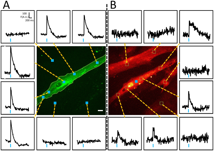
Excitation-contraction coupling in muscle cells is initiated by a restricted membrane depolarization delimited within the neuromuscular junction. This targeted depolarization triggers an action potential that propagates and induces a global cellular calcium response and a consequent contraction. To date, numerous studies have investigated this excitation-calcium response coupling by using different techniques to depolarize muscle cells. However, none of these techniques mimic the temporal and spatial resolution of membrane depolarization observed in the neuromuscular junction. By using optogenetics in C2C12 muscle cells, we developed a technique to study the calcium response following membrane depolarization induced by photostimulations of membrane surface similar or narrower than the neuromuscular junction area. These stimulations coupled to confocal calcium imaging generate a global cellular calcium response that is the consequence of a membrane depolarization propagation. In this context, this technique provides an interesting, contactless and relatively easy way of investigation of calcium increase/release as well as calcium decrease/re-uptake triggered by a propagated membrane depolarization.
肌细胞中的兴奋-收缩偶联是由神经肌肉接头内限定的受限膜去极化引发的。这种靶向去极化引发动作电位传播,并诱导全局细胞钙反应和随后的收缩。迄今为止,许多研究已经使用不同的技术来使肌细胞去极化来研究这种兴奋-钙反应偶联。然而,这些技术都无法模拟在神经肌肉接头中观察到的膜去极化的时间和空间分辨率。通过在 C2C12 肌细胞中使用光遗传学,我们开发了一种技术,用于研究通过光刺激类似于或小于神经肌肉接头区域的膜表面诱导的膜去极化后的钙反应。这些刺激与共聚焦钙成像相结合,产生全局细胞钙反应,这是膜去极化传播的结果。在这种情况下,该技术提供了一种有趣的、非接触式且相对简单的方法,用于研究由传播的膜去极化引发的钙增加/释放以及钙减少/再摄取。