Mitsuno Daisuke, Ueda Koichi, Itamiya Tomoki, Nuri Takashi, Otsuki Yuki
Department of Plastic and Reconstructive Surgery, Osaka Medical College, Osaka, Japan; and Department of Media Informatics, Aichi University of Technology, Aichi, Japan.
Plast Reconstr Surg Glob Open. 2017 Aug 2;5(8):e1432. doi: 10.1097/GOX.0000000000001432. eCollection 2017 Aug.
Augmented reality (AR) technology that can combine computer-generated images with a real scene has been reported in the medical field recently. We devised the AR system for evaluation of improvements of the body surface, which is important for plastic surgery.
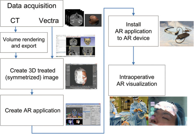
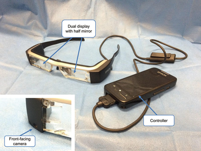
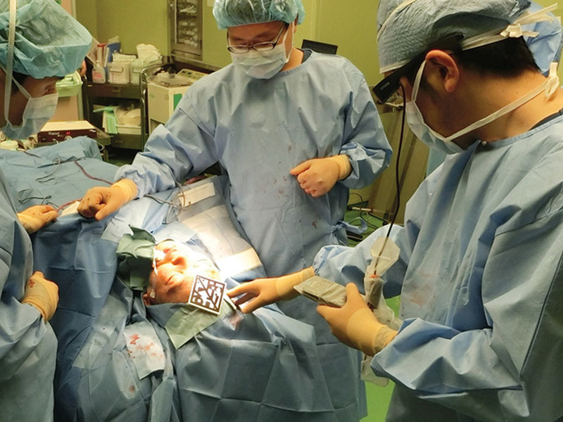
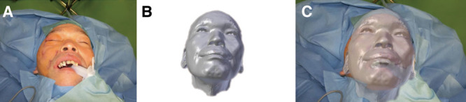
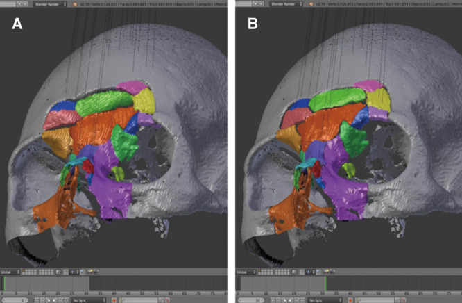
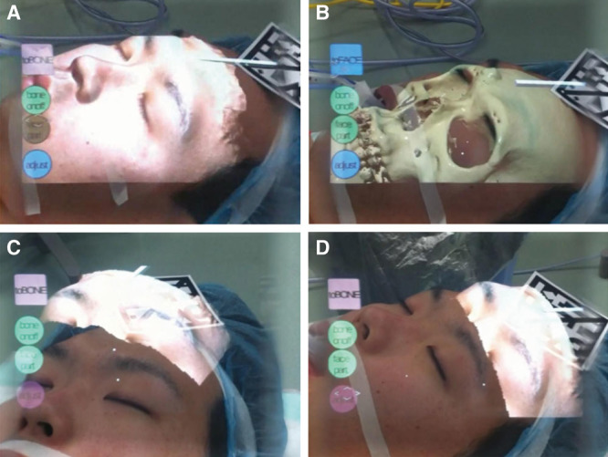
We constructed an AR system that is easy to modify by combining existing devices and free software. We superimposed the 3-dimensional images of the body surface and the bone (obtained from VECTRA H1 and CT) onto the actual surgical field by Moverio BT-200 smart glasses and evaluated improvements of the body surface in 8 cases.
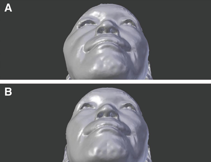
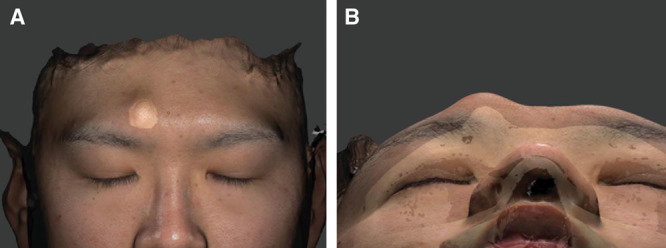
In all cases, the 3D image was successfully projected on the surgical field. Improvement of the display method of the 3D image made it easier to distinguish the different shapes in the 3D image and surgical field, making comparison easier. In a patient with fibrous dysplasia, the symmetrized body surface image was useful for confirming improvement of the real body surface. In a patient with complex facial fracture, the simulated bone image was useful as a reference for reduction. In a patient with an osteoma of the forehead, simultaneously displayed images of the body surface and the bone made it easier to understand these positional relationships.
This study confirmed that AR technology is helpful for evaluation of the body surface in several clinical applications. Our findings are not only useful for body surface evaluation but also for effective utilization of AR technology in the field of plastic surgery.
最近在医学领域报道了一种能将计算机生成的图像与真实场景相结合的增强现实(AR)技术。我们设计了用于评估体表改善情况的AR系统,这对整形手术很重要。
我们通过结合现有设备和免费软件构建了一个易于修改的AR系统。我们通过Moverio BT - 200智能眼镜将体表和骨骼的三维图像(从VECTRA H1和CT获得)叠加到实际手术区域,并评估了8例患者的体表改善情况。
在所有病例中,三维图像均成功投射到手术区域。三维图像显示方法的改进使得在三维图像和手术区域中更容易区分不同形状,便于比较。在一名患有骨纤维异常增殖症的患者中,对称的体表图像有助于确认真实体表的改善情况。在一名患有复杂面部骨折的患者中,模拟的骨骼图像有助于作为复位的参考。在一名患有前额骨瘤的患者中,同时显示的体表和骨骼图像使更容易理解这些位置关系。
本研究证实AR技术在多种临床应用中有助于评估体表。我们的研究结果不仅对体表评估有用,而且对AR技术在整形手术领域的有效利用也有用。