Memorial Sloan Kettering Cancer Center , New York, New York 10065, United States.
Department of Chemical Engineering, University of Rhode Island , Kingston, Rhode Island 02881, United States.
ACS Nano. 2017 Nov 28;11(11):10689-10703. doi: 10.1021/acsnano.7b04743. Epub 2017 Sep 12.
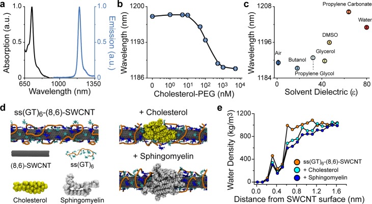
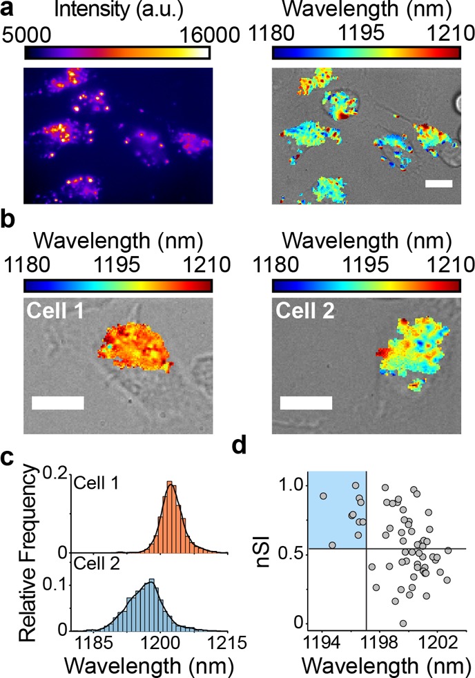
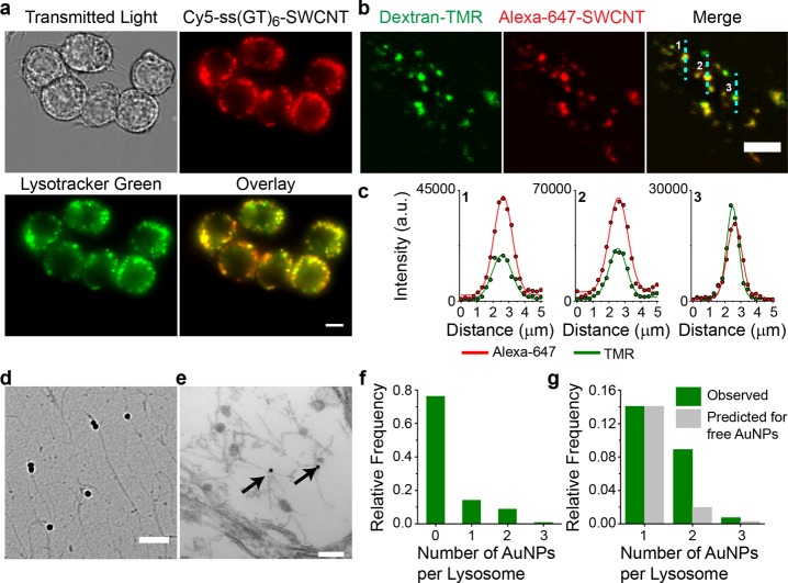
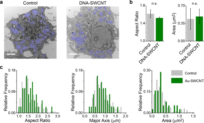
Lipid accumulation within the lumen of endolysosomal vesicles is observed in various pathologies including atherosclerosis, liver disease, neurological disorders, lysosomal storage disorders, and cancer. Current methods cannot measure lipid flux specifically within the lysosomal lumen of live cells. We developed an optical reporter, composed of a photoluminescent carbon nanotube of a single chirality, that responds to lipid accumulation via modulation of the nanotube's optical band gap. The engineered nanomaterial, composed of short, single-stranded DNA and a single nanotube chirality, localizes exclusively to the lumen of endolysosomal organelles without adversely affecting cell viability or proliferation or organelle morphology, integrity, or function. The emission wavelength of the reporter can be spatially resolved from within the endolysosomal lumen to generate quantitative maps of lipid content in live cells. Endolysosomal lipid accumulation in cell lines, an example of drug-induced phospholipidosis, was observed for multiple drugs in macrophages, and measurements of patient-derived Niemann-Pick type C fibroblasts identified lipid accumulation and phenotypic reversal of this lysosomal storage disease. Single-cell measurements using the reporter discerned subcellular differences in equilibrium lipid content, illuminating significant intracellular heterogeneity among endolysosomal organelles of differentiating bone-marrow-derived monocytes. Single-cell kinetics of lipoprotein-derived cholesterol accumulation within macrophages revealed rates that differed among cells by an order of magnitude. This carbon nanotube optical reporter of endolysosomal lipid content in live cells confers additional capabilities for drug development processes and the investigation of lipid-linked diseases.
在内体溶酶体囊泡的腔室内发生的脂质积累,可见于多种病理学中,包括动脉粥样硬化、肝脏疾病、神经紊乱、溶酶体贮积病和癌症。目前的方法无法特异性测量活细胞内溶酶体腔室内的脂质流量。我们开发了一种光学报告子,由单一手性的发光明细管碳纳米管组成,通过纳米管光学带隙的调制来响应脂质积累。这种工程纳米材料由短的单链 DNA 和单一的纳米管手性组成,特异性定位于内体溶酶体细胞器的腔室内,而不会对细胞活力、增殖或细胞器形态、完整性或功能产生不利影响。报告子的发射波长可以在腔室内从空间上分辨出来,从而在活细胞中生成脂质含量的定量图谱。我们观察到细胞系中的内体溶酶体脂质积累,这是药物诱导的磷脂沉积的一个例子,在巨噬细胞中观察到多种药物的内体溶酶体脂质积累,并对来源于尼曼-匹克 C 型患者的成纤维细胞进行了测量,鉴定了这种溶酶体贮积病的脂质积累和表型逆转。使用报告子的单细胞测量能够辨别出平衡脂质含量的亚细胞差异,揭示了分化的骨髓来源单核细胞中的内体溶酶体细胞器之间存在显著的细胞内异质性。巨噬细胞中脂蛋白衍生胆固醇积累的单细胞动力学显示,细胞之间的速率差异达到了数量级。这种活细胞内体溶酶体脂质含量的碳纳米管光学报告子为药物开发过程和脂质相关疾病的研究提供了额外的功能。