Bai Zhibin, Shi Yaoping, Wang Jianfeng, Qiu Longhua, Teng Gaojun, Zhang Feng, Yang Xiaoming
Image-Guided Biomolecular Intervention Research, Section of Interventional Radiology, Department of Radiology, University of Washington School of Medicine, Seattle, WA, USA.
Department of Radiology, Zhongda Hospital, Southeastern University, Nanjing, China.
Oncotarget. 2017 Apr 21;8(33):54277-54284. doi: 10.18632/oncotarget.17347. eCollection 2017 Aug 15.
To investigate the feasibility of using multi-modality imaging to monitor the creation of rat models with orthotopic pancreatic head cancer with obstructive jaundice.
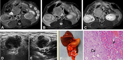
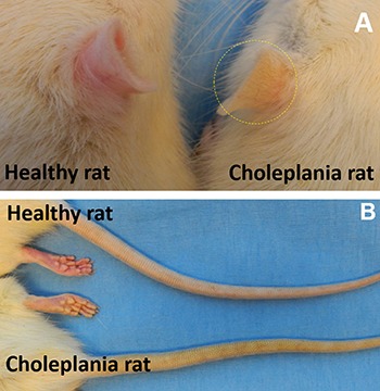
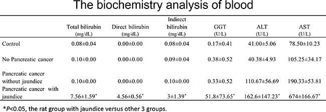
27 of 52 rats (51.92%) developed pancreatic head cancer. The tumor formation rate was significantly higher in the animal group receiving bioluminescent tumor, compared to the group receiving non-bioluminescent donor tumors [78.1% (25/32 rats) vs 10.0% (2/20 rats), = 0.0001]. Both ultrasound imaging and MRI clearly characterized the orthotopic tumors. Laboratory biochemistry test for those rats with obstructive jaundice showed elevated levels of bilirubin, aspartate transaminase (AST), alkaline phosphatase (ALT) and gamma-glutamyl transpeptidase (λ-GGT), compared with those rats without jaundice ( < 0.05). Correlative pathology confirmed that all tumors were ductal adenocarcinomas, and located in pancreatic head regions.
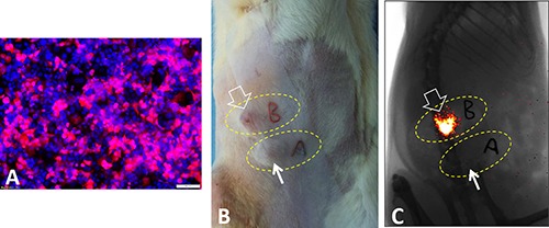
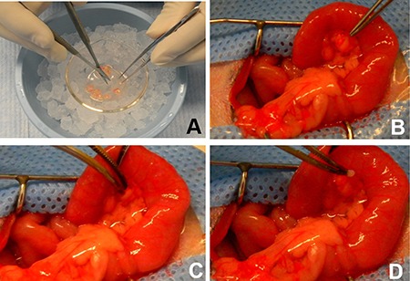
Rat pancreatic adenocarcinoma cells (DSL-6A/C1) were first transfected with lentivirus/mCherry-luciferase genes, and then subcutaneously implanted into flanks of donor immunocompetent Lewis rats, to create pancreatic tumor tissues. The tumor tissues from donor rats with either bioluminescence signal or without the signal were then transplanted into the pancreatic heads of 52 recipient Lewis rats. Bioluminescence optical and ultrasound imaging, as well as magnetic resonance imaging (MRI), were performed to follow up the tumor formation and growth in these tumor-transplanted rats. Physical examination and biochemistry test were used to discern the rats with obstructive jaundice. The rats were euthanized for subsequent histologic correlation and confirmation.
We successfully created a new rat model with orthotopic pancreatic head cancer, which can be accurately monitored and visualized by different imaging modalities.
探讨使用多模态成像监测伴有梗阻性黄疸的原位胰头癌大鼠模型构建的可行性。
52只大鼠中有27只(51.92%)发生了胰头癌。与接受无生物发光供体肿瘤的组相比,接受生物发光肿瘤的动物组肿瘤形成率显著更高[78.1%(25/32只大鼠)对10.0%(2/20只大鼠),P = 0.0001]。超声成像和磁共振成像(MRI)均能清晰地显示原位肿瘤的特征。与无黄疸的大鼠相比,那些伴有梗阻性黄疸的大鼠的实验室生化检测显示胆红素、天冬氨酸转氨酶(AST)、碱性磷酸酶(ALT)和γ-谷氨酰转肽酶(λ-GGT)水平升高(P < 0.05)。相关病理学证实所有肿瘤均为导管腺癌,且位于胰头区域。
首先用慢病毒/红色荧光蛋白-荧光素酶基因转染大鼠胰腺腺癌细胞(DSL-6A/C1),然后将其皮下植入具有免疫活性的供体Lewis大鼠的侧腹,以构建胰腺肿瘤组织。然后将来自有或无生物发光信号的供体大鼠的肿瘤组织移植到52只受体Lewis大鼠的胰头。采用生物发光光学成像、超声成像以及磁共振成像(MRI)对这些肿瘤移植大鼠的肿瘤形成和生长进行随访。通过体格检查和生化检测来识别伴有梗阻性黄疸的大鼠。对大鼠实施安乐死以进行后续的组织学关联和确认。
我们成功构建了一种新的原位胰头癌大鼠模型,该模型可通过不同的成像方式进行准确监测和可视化。