Ashok Iyaswamy, Sheeladevi Rathinasamy
Department of Physiology, Dr. ALM PG Institute of Basic Medical Sciences, University of Madras, Chennai, India.
J Food Drug Anal. 2015 Dec;23(4):679-691. doi: 10.1016/j.jfda.2014.07.011. Epub 2015 Jan 9.
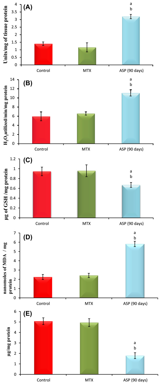
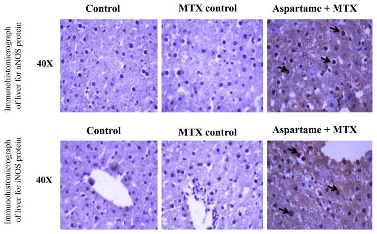
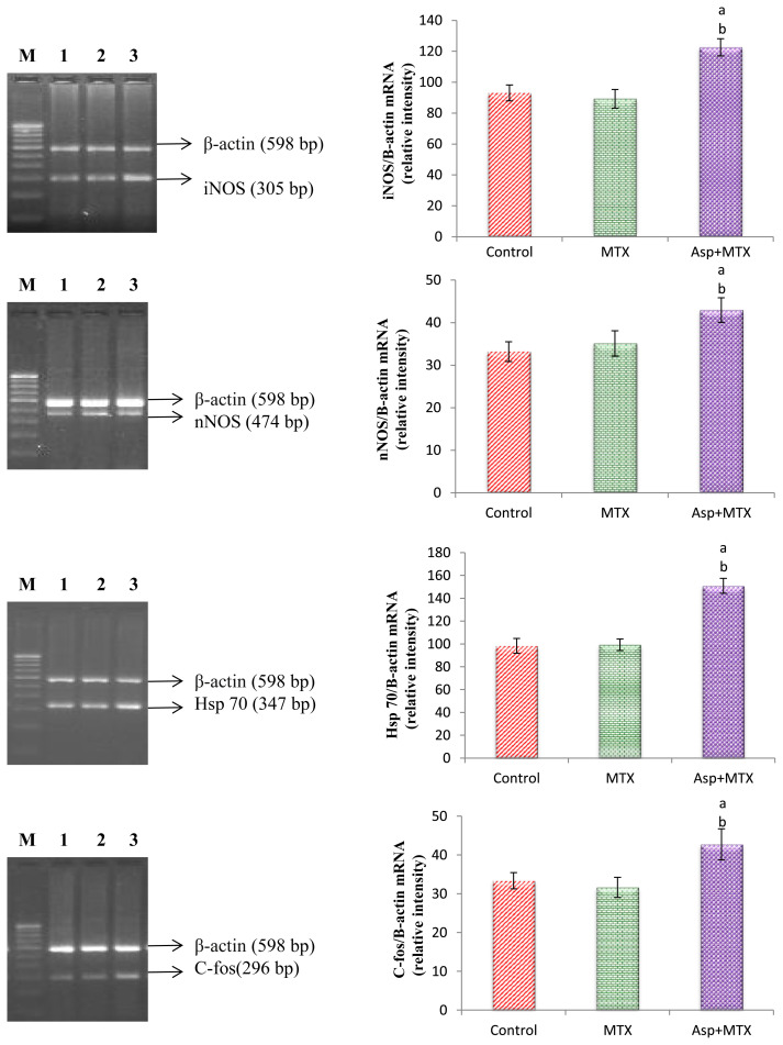
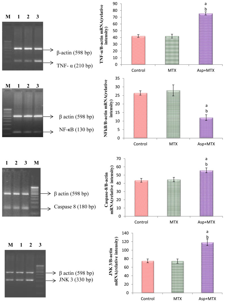
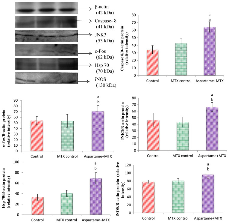
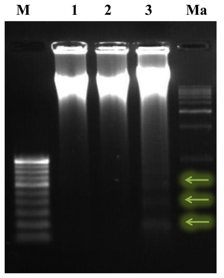
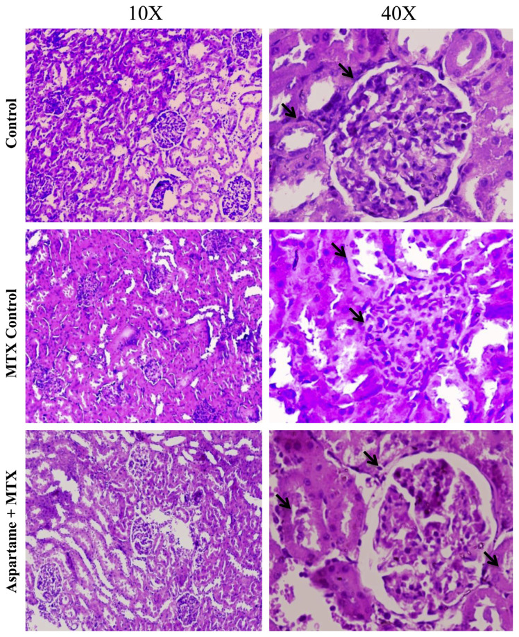
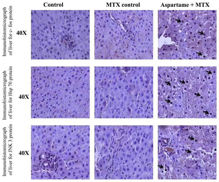
This study investigates how long-term (40 mg/kg b.wt) consumption of aspartame can alter the antioxidant status, stress pathway genes, and apoptotic changes in the liver of Wistar albino rats. Numerous controversial reports are available on the use of aspartame as it releases methanol as one of its metabolites during metabolism. To mimic the human methanol metabolism the methotrexate treated rats were included to study the aspartame effects. The aspartame treated methotrexate (MTX animals showed a marked significant increase in the superoxide dismutase (SOD), catalase (CAT), lipid peroxidation (LPO), and Glutathione peroxidase (GPx) activity in the liver from control and MTX control animals, and showed a significant decrease in reduced glutathione (GSH) and protein thiol in aspartame treated animals. The aspartame treated MTX animals showed a marked significant decrease in the body weight, brain, and liver weight. The aspartame treated MTX animals showed a marked increase in the inducible nitric oxide (iNOS), neuronal nitric oxide (nNOS), c-fos, Heat shock protein (Hsp) 70 Tumour necrosis Factor (TNF)α, caspase 8, c-jun N terminal kinases (JNK) 3 and Nuclear factor kappa B (NFkB) gene expression in the liver from control and MTX control animals. The aspartame treated MTX animals showed a marked increase in the c-fos, Hsp 70, iNOS Caspase 8, and JNK 3 protein expression in the liver from control and MTX control animals indicating the enhancement of stress and apoptosis. The aspartame treated MTX animals showed a streak of marked DNA fragmentation in the liver. On immunohistochemical analysis aspartame treated animals showed brown colored positive hepatocytes indicating the stress specific and apoptotic protein expression. Since aspartame consumption is on the rise among people, it is essential to create awareness regarding the usage of this artificial sweetener.
本研究调查了长期(40毫克/千克体重)摄入阿斯巴甜如何改变Wistar白化大鼠肝脏中的抗氧化状态、应激途径基因和凋亡变化。关于阿斯巴甜的使用有许多有争议的报道,因为它在代谢过程中会释放甲醇作为其代谢产物之一。为了模拟人类的甲醇代谢,纳入了用甲氨蝶呤处理的大鼠来研究阿斯巴甜的影响。用阿斯巴甜处理的甲氨蝶呤(MTX)动物肝脏中的超氧化物歧化酶(SOD)、过氧化氢酶(CAT)、脂质过氧化(LPO)和谷胱甘肽过氧化物酶(GPx)活性,与对照和MTX对照动物相比有显著明显增加,而在阿斯巴甜处理的动物中还原型谷胱甘肽(GSH)和蛋白质硫醇显著减少。用阿斯巴甜处理的MTX动物的体重、脑重和肝重显著明显下降。用阿斯巴甜处理的MTX动物肝脏中诱导型一氧化氮合酶(iNOS)、神经元型一氧化氮合酶(nNOS)、c-fos、热休克蛋白(Hsp)70、肿瘤坏死因子(TNF)α、半胱天冬酶8、c-jun氨基末端激酶(JNK)3和核因子κB(NFkB)基因表达,与对照和MTX对照动物相比有显著明显增加。用阿斯巴甜处理的MTX动物肝脏中c-fos、Hsp 70、iNOS、半胱天冬酶8和JNK 3蛋白表达显著明显增加,表明应激和凋亡增强。用阿斯巴甜处理的MTX动物肝脏中有明显的DNA片段化条纹。免疫组织化学分析显示,用阿斯巴甜处理的动物肝脏中有棕色阳性肝细胞,表明有应激特异性和凋亡蛋白表达。由于阿斯巴甜的消费量在人群中不断上升,因此有必要提高对这种人工甜味剂使用的认识。