Puri Tanuj, Greenhalgh Tessa A, Wilson James M, Franklin Jamie, Wang Lia Mun, Strauss Victoria, Cunningham Chris, Partridge Mike, Maughan Tim
CRUK/MRC Oxford Institute of Radiation Oncology, Department of Oncology, University of Oxford, Old Road Campus Research Building, Off Roosevelt Drive, Oxford, OX3 7DQ, UK.
Department of Radiology, Oxford University Hospitals NHS Foundation Trust, Oxford, UK.
EJNMMI Res. 2017 Sep 20;7(1):78. doi: 10.1186/s13550-017-0324-x.
There is an increasing interest in developing predictive biomarkers of tissue hypoxia using functional imaging for personalised radiotherapy in patients with rectal cancer that are considered for neoadjuvant chemoradiotherapy (CRT). The study explores [F]fluoromisonidazole ([F]FMISO) positron emission tomography (PET) scans for predicting clinical response in rectal cancer patients receiving neoadjuvant CRT.
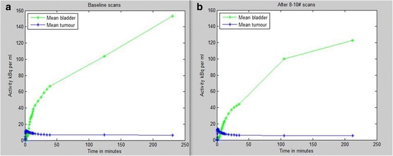
Patients with biopsy-proven rectal adenocarcinoma were imaged at 0-45 min, 2 and 4 h, at baseline and after 8-10 fractions of CRT (week 2). The first 6 patients did not receive an enema (the non-enema group) and the last 4 patients received an enema before PET-CT scan (the enema group). [F]FMISO production failed on 2 occasions. Static PET images at 4 h were analysed using tumour-to-muscle (T:M) SUVmax and tumour-to-blood (T:B) SUVmax. The 0-45 min dynamic PET scans were analysed using Casciari model to report hypoxia and perfusion. Akaike information criteria (AIC) were used to compare data fittings for different pharmacokinetic models. Pathological tumour regression grade was scored using American Joint Committee on Cancer (AJCC) 7.0. Shapiro-Wilk test was used to evaluate the normality of the data.
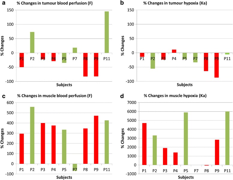
Five out of eleven (5/11) patients were classed as good responders (AJCC 0/1 or good clinical response) and 6/11 as poor responders (AJCC 2/3 or poor clinical response). The median T:M SUVmax was 2.14 (IQR 0.58) at baseline and 1.30 (IQR 0.19) at week 2, and the corresponding median tumour hypoxia volume was 1.08 (IQR 1.31) cm and 0 (IQR 0.15) cm, respectively. The median T:B SUVmax was 2.46 (IQR 1.50) at baseline and 1.61 (IQR 0.14) at week 2, and the corresponding median tumour hypoxia volume was 5.68 (IQR 5.86) cm and 0.76 (IQR 0.78) cm, respectively. For 0-45 min tumour modelling, the median hypoxia was 0.92 (IQR 0.41) min at baseline and 0.70 (IQR 0.10) min at week 2. The median perfusion was 4.10 (IQR 1.71) ml g min at baseline and 2.48 (IQR 3.62) ml g min at week 2. In 9/11 patients with both PET scans, tumour perfusion decreased in non-responders and increased in responders except in one patient. None of the changes in other PET parameters showed any clear trend with clinical outcome.
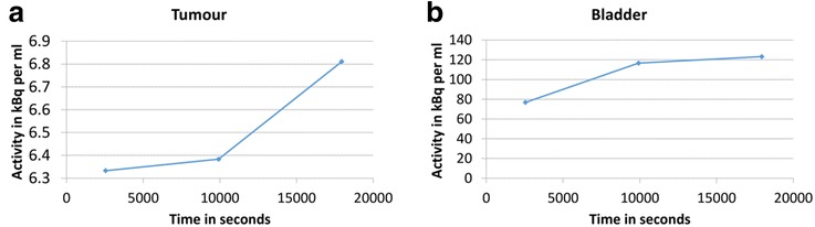
This pilot study with small number of datasets revealed significant challenges in delivery and interpretation of [F]FMISO PET scans of rectal cancer. There are two principal problems namely spill-in from non-tumour tracer activity from rectal and bladder contents. Emphasis should be made on reducing spill-in effects from the bladder to improve data quality. This preliminary study has shown fundamental difficulties in the interpretation of [F]FMISO PET scans for rectal cancer, limiting its clinical applicability.
利用功能成像技术开发组织缺氧的预测生物标志物,用于考虑新辅助放化疗(CRT)的直肠癌患者的个体化放疗,这一兴趣日益浓厚。本研究探讨[F]氟米索硝唑([F]FMISO)正电子发射断层扫描(PET)在预测接受新辅助CRT的直肠癌患者临床反应中的应用。
经活检证实为直肠腺癌的患者在基线时、CRT的8 - 10次分割后(第2周)的0 - 45分钟、2小时和4小时进行成像。前6例患者未接受灌肠(非灌肠组),后4例患者在PET - CT扫描前接受灌肠(灌肠组)。[F]FMISO制备失败2次。使用肿瘤与肌肉(T:M)SUVmax和肿瘤与血液(T:B)SUVmax分析4小时的静态PET图像。使用Casciari模型分析0 - 45分钟的动态PET扫描以报告缺氧和灌注情况。使用赤池信息准则(AIC)比较不同药代动力学模型的数据拟合情况。使用美国癌症联合委员会(AJCC)7.0对病理肿瘤退缩分级进行评分。使用Shapiro - Wilk检验评估数据的正态性。
11例患者中有5例(5/11)被归类为良好反应者(AJCC 0/1或良好临床反应),6/11为不良反应者(AJCC 2/3或不良临床反应)。基线时T:M SUVmax的中位数为2.14(IQR 0.58),第2周时为1.30(IQR 0.19),相应的肿瘤缺氧体积中位数分别为1.08(IQR 1.31)cm和0(IQR 0.15)cm。基线时T:B SUVmax的中位数为2.46(IQR 1.50),第2周时为1.61(IQR 0.14),相应的肿瘤缺氧体积中位数分别为5.68(IQR 5.86)cm和0.76(IQR 0.78)cm。对于0 - 45分钟的肿瘤建模,基线时缺氧的中位数为0.92(IQR 0.41)分钟,第2周时为0.70(IQR 0.10)分钟。基线时灌注的中位数为4.10(IQR 1.71)ml g min,第2周时为2.48(IQR 3.62)ml g min。在11例进行了两次PET扫描的患者中,除1例患者外,无反应者的肿瘤灌注降低,反应者的肿瘤灌注增加。其他PET参数的变化均未显示出与临床结果有任何明显趋势。
这项包含少量数据集的初步研究揭示了直肠癌[F]FMISO PET扫描在实施和解读方面存在重大挑战。存在两个主要问题,即来自直肠和膀胱内容物的非肿瘤示踪剂活性的溢出。应着重减少来自膀胱的溢出效应以提高数据质量。这项初步研究表明在直肠癌[F]FMISO PET扫描的解读方面存在根本困难,限制了其临床应用。