• 文献检索
  • 文档翻译
  • 深度研究
  • 学术资讯
  • Suppr Zotero 插件Zotero 插件
  • 邀请有礼
  • 套餐&价格
  • 历史记录
应用&插件
Suppr Zotero 插件Zotero 插件浏览器插件Mac 客户端Windows 客户端微信小程序
定价
高级版会员购买积分包购买API积分包
服务
文献检索文档翻译深度研究API 文档MCP 服务
关于我们
关于 Suppr公司介绍联系我们用户协议隐私条款
关注我们

Suppr 超能文献

核心技术专利:CN118964589B侵权必究
粤ICP备2023148730 号-1Suppr @ 2026

文献检索

告别复杂PubMed语法,用中文像聊天一样搜索,搜遍4000万医学文献。AI智能推荐,让科研检索更轻松。

立即免费搜索

文件翻译

保留排版,准确专业,支持PDF/Word/PPT等文件格式,支持 12+语言互译。

免费翻译文档

深度研究

AI帮你快速写综述,25分钟生成高质量综述,智能提取关键信息,辅助科研写作。

立即免费体验

用于抑制正电子发射断层扫描中高放射性区域附近溢出效应的生理背景校正方法的验证

Validation of the physiological background correction method for the suppression of the spill-in effect near highly radioactive regions in positron emission tomography.

作者信息

Akerele Mercy I, Wadhwa Palak, Silva-Rodriguez Jesus, Hallett William, Tsoumpas Charalampos

机构信息

Biomedical Imaging Science Department, School of Medicine, University of Leeds, Leeds, West Yorkshire, UK.

Invicro, Hammersmith Hospital, London, UK.

出版信息

EJNMMI Phys. 2018 Dec 5;5(1):34. doi: 10.1186/s40658-018-0233-8.

DOI:10.1186/s40658-018-0233-8
PMID:30519974
原文链接:https://pmc.ncbi.nlm.nih.gov/articles/PMC6281548/
Abstract

BACKGROUND

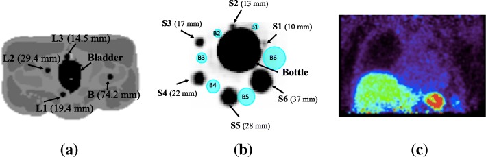
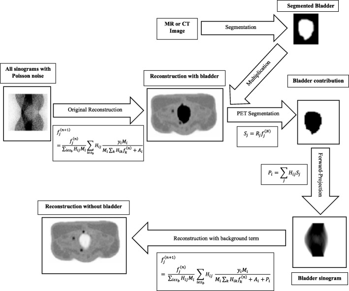
Positron emission tomography (PET) imaging has a wide applicability in oncology, cardiology and neurology. However, a major drawback when imaging very active regions such as the bladder is the spill-in effect, leading to inaccurate quantification and obscured visualisation of nearby lesions. Therefore, this study aims at investigating and correcting for the spill-in effect from high-activity regions to the surroundings as a function of activity in the hot region, lesion size and location, system resolution and application of post-filtering using a recently proposed background correction technique. This study involves analytical simulations for the digital XCAT2 phantom and validation acquiring NEMA phantom and patient data with the GE Signa PET/MR scanner. Reconstructions were done using the ordered subset expectation maximisation (OSEM) algorithm. Dedicated point-spread function (OSEM+PSF) and a recently proposed background correction (OSEM+PSF+BC) were incorporated into the reconstruction for spill-in correction. The standardised uptake values (SUV) were compared for all reconstruction algorithms.

RESULTS

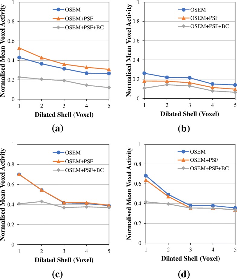
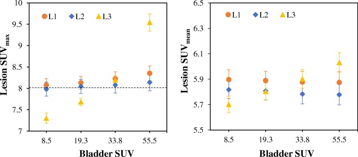
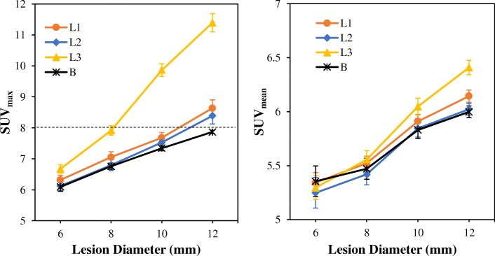
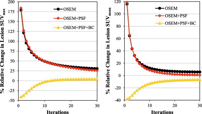
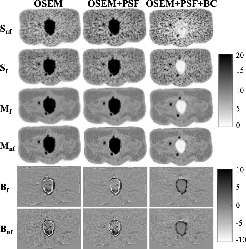
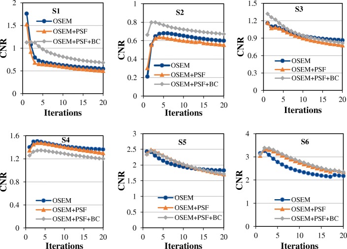
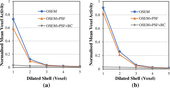
The simulation study revealed that lesions within 15-20 mm from the hot region were predominantly affected by the spill-in effect, leading to an increased bias and impaired lesion visualisation within the region. For OSEM, lesion SUV converged to the true value at low bladder activity, but as activity increased, there was an overestimation as much as 19% for proximal lesions (distance around 15-20 mm from the bladder edge) and 2-4% for distant lesions (distance larger than 20 mm from the bladder edge). As bladder SUV increases, the % SUV change for proximal lesions is about 31% and 6% for SUV and SUV, respectively, showing that the spill-in effect is more evident for the SUV than the SUV. Also, the application of post-filtering resulted in up to 65% increment in the spill-in effect around the bladder edges. For proximal lesions, PSF has no major improvement over OSEM because of the spill-in effect, coupled with the blurring effect by post-filtering. Within two voxels around the bladder, the spill-in effect in OSEM is 42% (32%), while for OSEM+PSF, it is 31% (19%), with (and without) post-filtering, respectively. But with OSEM+PSF+BC, the spill-in contribution from the bladder was relatively low (below 5%, either with or without post-filtering). These results were further validated using the NEMA phantom and patient data for which OSEM+PSF+BC showed about 70-80% spill-in reduction around the bladder edges and increased contrast-to-noise ratio up to 36% compared to OSEM and OSEM+PSF reconstructions without post-filtering.

CONCLUSION

The spill-in effect is dependent on the activity in the hot region, lesion size and location, as well as post-filtering; and this is more evident in SUV than SUV. However, the recently proposed background correction method facilitates stability in quantification and enhances the contrast in lesions with low uptake.

摘要

背景

正电子发射断层扫描(PET)成像在肿瘤学、心脏病学和神经病学领域具有广泛的适用性。然而,在对膀胱等非常活跃的区域进行成像时,一个主要缺点是溢出效应,这会导致定量不准确以及附近病变的可视化模糊。因此,本研究旨在研究并校正从高活性区域到周围环境的溢出效应,该效应是热区活性、病变大小和位置、系统分辨率以及使用最近提出的背景校正技术进行后滤波应用的函数。本研究涉及对数字XCAT2体模的分析模拟,以及使用GE Signa PET/MR扫描仪获取NEMA体模和患者数据进行验证。使用有序子集期望最大化(OSEM)算法进行重建。将专用点扩散函数(OSEM+PSF)和最近提出的背景校正(OSEM+PSF+BC)纳入重建以进行溢出校正。比较了所有重建算法的标准化摄取值(SUV)。

结果

模拟研究表明,距离热区15 - 20毫米内的病变主要受溢出效应影响,导致该区域内偏差增加且病变可视化受损。对于OSEM,在膀胱低活性时病变SUV收敛到真实值,但随着活性增加,近端病变(距离膀胱边缘约15 - 20毫米)的高估高达19%,远端病变(距离膀胱边缘大于20毫米)的高估为2 - 4%。随着膀胱SUV增加,近端病变的SUV变化百分比在SUV和SUV时分别约为31%和6%,表明溢出效应在SUV中比在SUV中更明显。此外,后滤波的应用导致膀胱边缘周围的溢出效应增加高达65%。对于近端病变,由于溢出效应以及后滤波的模糊效应,PSF相对于OSEM没有显著改善。在膀胱周围的两个体素内,OSEM中的溢出效应为42%(32%),而对于OSEM+PSF,分别为31%(19%),分别有(和没有)后滤波。但使用OSEM+PSF+BC时,来自膀胱的溢出贡献相对较低(无论有无后滤波均低于5%)。使用NEMA体模和患者数据进一步验证了这些结果,与未进行后滤波的OSEM和OSEM+PSF重建相比,OSEM+PSF+BC在膀胱边缘周围显示出约70 - 80%的溢出减少,并且对比度噪声比提高了36%。

结论

溢出效应取决于热区活性、病变大小和位置以及后滤波;并且在SUV中比在SUV中更明显。然而,最近提出的背景校正方法有助于定量的稳定性并增强低摄取病变的对比度。

https://cdn.ncbi.nlm.nih.gov/pmc/blobs/0357/6281548/e201cbe45258/40658_2018_233_Fig12_HTML.jpg
https://cdn.ncbi.nlm.nih.gov/pmc/blobs/0357/6281548/6996c68bc3cc/40658_2018_233_Fig1_HTML.jpg
https://cdn.ncbi.nlm.nih.gov/pmc/blobs/0357/6281548/2d541dd78892/40658_2018_233_Fig2_HTML.jpg
https://cdn.ncbi.nlm.nih.gov/pmc/blobs/0357/6281548/8f06389572f8/40658_2018_233_Fig3_HTML.jpg
https://cdn.ncbi.nlm.nih.gov/pmc/blobs/0357/6281548/c68f8da9a425/40658_2018_233_Fig4_HTML.jpg
https://cdn.ncbi.nlm.nih.gov/pmc/blobs/0357/6281548/b96c0e4a408f/40658_2018_233_Fig5_HTML.jpg
https://cdn.ncbi.nlm.nih.gov/pmc/blobs/0357/6281548/c421b75645f3/40658_2018_233_Fig6_HTML.jpg
https://cdn.ncbi.nlm.nih.gov/pmc/blobs/0357/6281548/25cb1ffea269/40658_2018_233_Fig7_HTML.jpg
https://cdn.ncbi.nlm.nih.gov/pmc/blobs/0357/6281548/e74b857f8b89/40658_2018_233_Fig8_HTML.jpg
https://cdn.ncbi.nlm.nih.gov/pmc/blobs/0357/6281548/83cea27c8840/40658_2018_233_Fig9_HTML.jpg
https://cdn.ncbi.nlm.nih.gov/pmc/blobs/0357/6281548/c20cecbcc920/40658_2018_233_Fig10_HTML.jpg
https://cdn.ncbi.nlm.nih.gov/pmc/blobs/0357/6281548/2d5e95dbc06d/40658_2018_233_Fig11_HTML.jpg
https://cdn.ncbi.nlm.nih.gov/pmc/blobs/0357/6281548/e201cbe45258/40658_2018_233_Fig12_HTML.jpg

相似文献

1
Validation of the physiological background correction method for the suppression of the spill-in effect near highly radioactive regions in positron emission tomography.用于抑制正电子发射断层扫描中高放射性区域附近溢出效应的生理背景校正方法的验证
EJNMMI Phys. 2018 Dec 5;5(1):34. doi: 10.1186/s40658-018-0233-8.
2
Impact of point spread function modelling and time of flight on FDG uptake measurements in lung lesions using alternative filtering strategies.使用替代滤波策略时,点扩散函数建模和飞行时间对肺部病变中FDG摄取测量的影响。
EJNMMI Phys. 2014 Dec;1(1):99. doi: 10.1186/s40658-014-0099-3. Epub 2014 Nov 30.
3
Clinical evaluation of whole-body oncologic PET with time-of-flight and point-spread function for the hybrid PET/MR system.用于PET/MR混合系统的具有飞行时间和点扩散函数的全身肿瘤PET的临床评估。
Eur J Radiol. 2017 Aug;93:70-75. doi: 10.1016/j.ejrad.2017.05.029. Epub 2017 May 25.
4
Iterative reconstruction incorporating background correction improves quantification of [F]-NaF PET/CT images of patients with abdominal aortic aneurysm.迭代重建结合背景校正可改善腹主动脉瘤患者 [F]-NaF PET/CT 图像的定量分析。
J Nucl Cardiol. 2021 Oct;28(5):1875-1886. doi: 10.1007/s12350-019-01940-4. Epub 2019 Nov 11.
5
The influence of different signal-to-background ratios on spatial resolution and F18-FDG-PET quantification using point spread function and time-of-flight reconstruction.不同信背比对使用点扩散函数和飞行时间重建的空间分辨率及F18-FDG-PET定量分析的影响。
EJNMMI Phys. 2014 Dec;1(1):12. doi: 10.1186/2197-7364-1-12. Epub 2014 Sep 19.
6
F-FDG PET/CT heterogeneity quantification through textural features in the era of harmonisation programs: a focus on lung cancer.在标准化计划时代通过纹理特征进行F-FDG PET/CT异质性量化:以肺癌为重点
Eur J Nucl Med Mol Imaging. 2016 Dec;43(13):2324-2335. doi: 10.1007/s00259-016-3441-2. Epub 2016 Jun 21.
7
The association of tumor-to-background ratios and SUVmax deviations related to point spread function and time-of-flight F18-FDG-PET/CT reconstruction in colorectal liver metastases.肿瘤与背景比值与 SUVmax 偏差与点扩散函数和飞行时间 F18-FDG-PET/CT 重建的关系在结直肠癌肝转移中的应用。
EJNMMI Res. 2015 May 6;5:31. doi: 10.1186/s13550-015-0111-5. eCollection 2015.
8
Impact of time of flight and point spread function on quantitative parameters of lung lesions in F-FDG PET/CT.飞行时间和点扩散函数对 F-FDG PET/CT 中肺病变定量参数的影响。
BMC Med Imaging. 2021 Nov 13;21(1):169. doi: 10.1186/s12880-021-00699-w.
9
Effect of Tumor-Pixel Positioning on the Variability of SUV Measurements in PET Images.肿瘤像素定位对PET图像中SUV测量值变异性的影响。
Asia Ocean J Nucl Med Biol. 2023;11(1):71-81. doi: 10.22038/AOJNMB.2022.61623.1434.
10
Performance measurement of PSF modeling reconstruction (True X) on Siemens Biograph TruePoint TrueV PET/CT.在西门子Biograph TruePoint TrueV PET/CT上对PSF建模重建(真实X)的性能测量。
Ann Nucl Med. 2014 May;28(4):340-8. doi: 10.1007/s12149-014-0815-z. Epub 2014 Feb 7.

引用本文的文献

1
The Additional Role of F18-FDG PET/CT in Characterizing MRI-Diagnosed Tumor Deposits in Locally Advanced Rectal Cancer.F18-FDG PET/CT 在 MRI 诊断局部晚期直肠癌肿瘤沉积中的附加作用。
Tomography. 2024 Apr 22;10(4):632-642. doi: 10.3390/tomography10040048.
2
Quantifying sodium [F]fluoride uptake in abdominal aortic aneurysms.定量腹主动脉瘤中[F]氟化钠的摄取情况。
EJNMMI Res. 2022 Jun 6;12(1):33. doi: 10.1186/s13550-022-00904-z.
3
Cannonball appearance, a tricky yet underrated imaging manifestation of COVID-19.炮弹征,一种狡猾却被低估的 COVID-19 影像学表现。

本文引用的文献

1
F-Sodium Fluoride Uptake in Abdominal Aortic Aneurysms: The SoFIA Study.腹部主动脉瘤中 F-氟化钠摄取:SoFIA 研究。
J Am Coll Cardiol. 2018 Feb 6;71(5):513-523. doi: 10.1016/j.jacc.2017.11.053.
2
[F]Fluoromisonidazole PET in rectal cancer.[F]氟代米索硝唑正电子发射断层扫描在直肠癌中的应用
EJNMMI Res. 2017 Sep 20;7(1):78. doi: 10.1186/s13550-017-0324-x.
3
Impact of partial-volume correction in oncological PET studies: a systematic review and meta-analysis.肿瘤 PET 研究中部分容积校正的影响:系统评价和荟萃分析。
Emerg Radiol. 2022 Feb;29(1):35-39. doi: 10.1007/s10140-021-01997-0. Epub 2021 Nov 13.
4
Effect of COVID-19 on F-FDG PET/CT: Is There a Need to Consider COVID-19 Status Before Planning F-FDG PET/CT for Oncologic Evaluation?COVID-19 对 F-FDG PET/CT 的影响:在计划 F-FDG PET/CT 进行肿瘤评估之前,是否需要考虑 COVID-19 状态?
J Nucl Med Technol. 2021 Sep;49(3):284-285. doi: 10.2967/jnmt.121.262145. Epub 2021 Jul 9.
5
Comparison of Correction Techniques for the Spill in Effect in Emission Tomography.发射断层成像中效应溢出校正技术的比较
IEEE Trans Radiat Plasma Med Sci. 2020 Jul;4(4):422-432. doi: 10.1109/TRPMS.2020.2980443. Epub 2020 Mar 12.
6
Assessment of different quantification metrics of [F]-NaF PET/CT images of patients with abdominal aortic aneurysm.评估 [F]-NaF PET/CT 图像在腹主动脉瘤患者中的不同量化指标。
J Nucl Cardiol. 2022 Feb;29(1):251-261. doi: 10.1007/s12350-020-02220-2. Epub 2020 Jun 17.
7
Optimizing arterial F-sodium fluoride positron emission tomography analysis.优化动脉氟化钠正电子发射断层扫描分析。
J Nucl Cardiol. 2021 Oct;28(5):1887-1890. doi: 10.1007/s12350-019-01992-6. Epub 2019 Dec 23.
8
Spill-in counts in the quantification of F-florbetapir on Aβ-negative subjects: the effect of including white matter in the reference region.Aβ阴性受试者中F-氟代硼替佐米定量时的溢出计数:参考区域纳入白质的影响
EJNMMI Phys. 2019 Dec 19;6(1):27. doi: 10.1186/s40658-019-0258-7.
9
Iterative reconstruction incorporating background correction improves quantification of [F]-NaF PET/CT images of patients with abdominal aortic aneurysm.迭代重建结合背景校正可改善腹主动脉瘤患者 [F]-NaF PET/CT 图像的定量分析。
J Nucl Cardiol. 2021 Oct;28(5):1875-1886. doi: 10.1007/s12350-019-01940-4. Epub 2019 Nov 11.
Eur J Nucl Med Mol Imaging. 2017 Nov;44(12):2105-2116. doi: 10.1007/s00259-017-3775-4. Epub 2017 Aug 4.
4
Partial volume correction for improved PET quantification in F-NaF imaging of atherosclerotic plaques.用于改善动脉粥样硬化斑块 F-NaF 成像中 PET 定量的部分容积校正。
J Nucl Cardiol. 2018 Oct;25(5):1742-1756. doi: 10.1007/s12350-017-0778-2. Epub 2017 Feb 7.
5
Point-spread function reconstructed PET images of sub-centimeter lesions are not quantitative.亚厘米级病变的点扩散函数重建PET图像不具有定量性。
EJNMMI Phys. 2017 Dec;4(1):5. doi: 10.1186/s40658-016-0169-9. Epub 2017 Jan 13.
6
Improved diagnosis of pelvic lesions with dual-phase (18F)FDG-PET/CT.利用双期(18F)氟代脱氧葡萄糖正电子发射断层显像/计算机断层扫描(FDG-PET/CT)改善盆腔病变的诊断
Radiol Case Rep. 2015 Nov 6;6(1):479. doi: 10.2484/rcr.v6i1.479. eCollection 2011.
7
(68)Ga-PSMA ligand PET/CT in patients with prostate cancer: How we review and report.前列腺癌患者的(68)Ga-PSMA配体PET/CT:我们的评估与报告方式
Cancer Imaging. 2016 Jun 8;16(1):14. doi: 10.1186/s40644-016-0072-6.
8
NEMA NU 2-2012 performance studies for the SiPM-based ToF-PET component of the GE SIGNA PET/MR system.通用电气Signa PET/MR系统基于硅光电倍增管的飞行时间正电子发射断层显像组件的NEMA NU 2-2012性能研究。
Med Phys. 2016 May;43(5):2334. doi: 10.1118/1.4945416.
9
Design Features and Mutual Compatibility Studies of the Time-of-Flight PET Capable GE SIGNA PET/MR System.基于飞行时间技术的正电子发射断层扫描/磁共振成像系统的设计特点及相互兼容性研究。
IEEE Trans Med Imaging. 2016 Aug;35(8):1907-14. doi: 10.1109/TMI.2016.2537811. Epub 2016 Mar 9.
10
Recent developments in time-of-flight PET.近年来飞行时间 PET 的发展。
EJNMMI Phys. 2016 Dec;3(1):3. doi: 10.1186/s40658-016-0138-3. Epub 2016 Feb 16.