Li Bing, Urban Jill Pg, Yu Jing
Department of Orthopedics, Tianjin Hospital, Tianjin, China.
Department of Physiology, Anatomy and Genetics, University of Oxford, Oxford, UK.
Bone Res. 2017 Feb 21;5:16053. doi: 10.1038/boneres.2016.53. eCollection 2017.
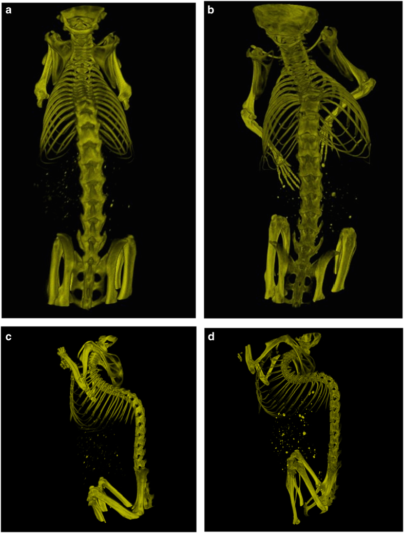
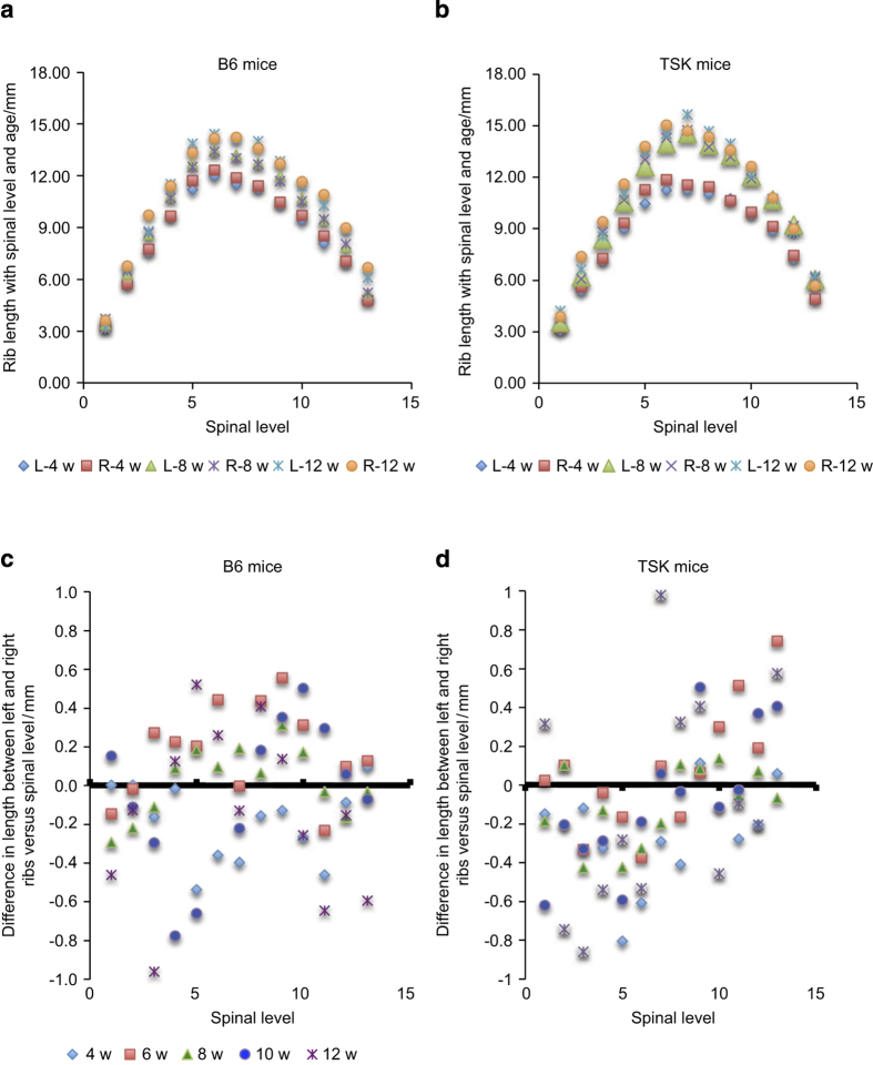
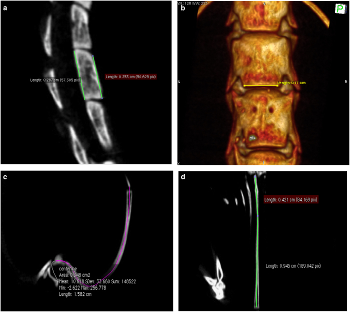
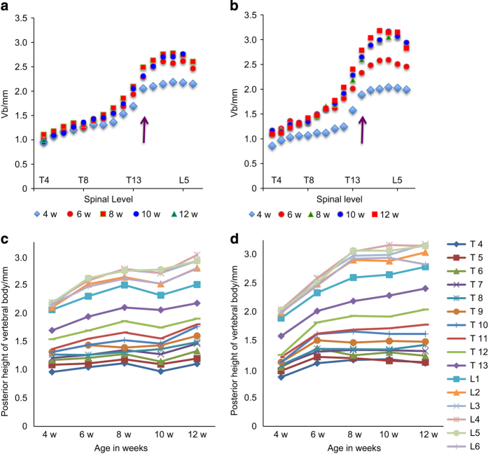
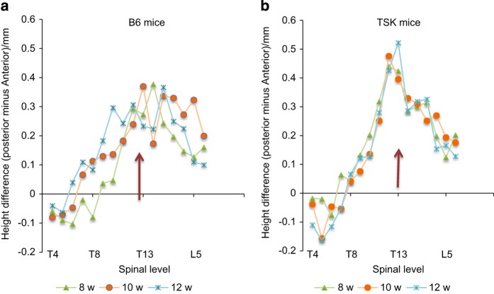
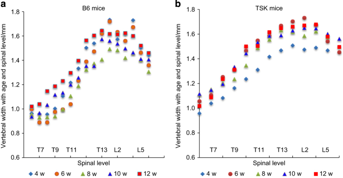
Tight-skin (TSK) mice are commonly used as an animal model to study the pathogenesis of Marfan syndrome (MFS), but little is known of their skeletal phenotype and in particular of the development of the spinal deformities, common in MFS. Here we examined growth of the axial skeletons of TSK and wild-type(B6) mice during their period of rapid growth. The whole bodies of mice, 4-12 weeks of age, were scanned after sacrifice, by micro-computed tomography (microCT). We reconstructed three-dimensional models of the spine and ribs, and measured vertebral body heights and rib lengths using the Mac-based image-processing software "OsiriX". Although the TSK mice were smaller than the B6 mice at 4 weeks, they experienced an early growth spurt and by 8 weeks the height, but not the width, of the vertebral body was significantly greater in the TSK mice than the B6 mice. Measurement of the angles of scoliotic and kyphotic curves post-mortem in the mice was problematic, hence we measured changes that develop in skeletal elements in these disorders. As a marker of kyphosis, we measured anterior wedging of the vertebral bodies; as a marker for scoliosis we measured asymmetries in rib length. We found, unlike in the B6 mice where the pattern was diffuse, wedging in TSK mice was directly related to spinal level and peaked steeply at the thoracolumbar junction. There was also significant asymmetry in length of the ribs in the TSK mice, but not in the B6 mice. The TSK mice thus appear to exhibit spinal deformities seen in MFS and could be a useful model for gaining understanding of the mechanisms of development of scoliosis and kyphosis in this disorder.
紧皮(TSK)小鼠通常被用作研究马凡综合征(MFS)发病机制的动物模型,但对其骨骼表型,尤其是MFS中常见的脊柱畸形的发展情况了解甚少。在此,我们研究了TSK小鼠和野生型(B6)小鼠在快速生长期间轴向骨骼的生长情况。对4至12周龄小鼠处死后的全身进行了微计算机断层扫描(microCT)。我们重建了脊柱和肋骨的三维模型,并使用基于Mac的图像处理软件“OsiriX”测量了椎体高度和肋骨长度。尽管4周龄时TSK小鼠比B6小鼠小,但它们经历了早期生长突增,到8周时,TSK小鼠椎体的高度而非宽度显著大于B6小鼠。在小鼠死后测量脊柱侧凸和后凸曲线的角度存在问题,因此我们测量了这些疾病中骨骼元素发生的变化。作为后凸的标志物,我们测量了椎体的前缘楔形变;作为脊柱侧凸的标志物,我们测量了肋骨长度的不对称性。我们发现,与B6小鼠中模式分散不同,TSK小鼠的楔形变与脊柱水平直接相关,并在胸腰段交界处急剧达到峰值。TSK小鼠的肋骨长度也存在显著不对称性,而B6小鼠则没有。因此,TSK小鼠似乎表现出MFS中出现的脊柱畸形,可能是了解该疾病中脊柱侧凸和后凸发展机制的有用模型。