Welton Joanne L, Loveless Samantha, Stone Timothy, von Ruhland Chris, Robertson Neil P, Clayton Aled
Department of Biomedical Sciences, Cardiff School of Health Sciences, Cardiff Metropolitan University, Cardiff, UK.
Division of Cancer and Genetics, School of Medicine, Cardiff University, Velindre Cancer Centre, Cardiff, UK.
J Extracell Vesicles. 2017 Sep 3;6(1):1369805. doi: 10.1080/20013078.2017.1369805. eCollection 2017.
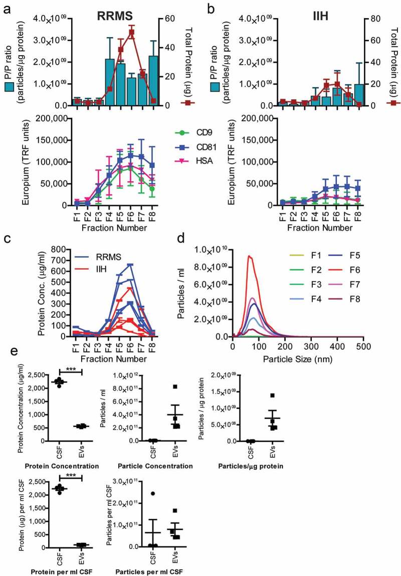
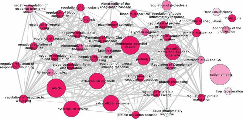
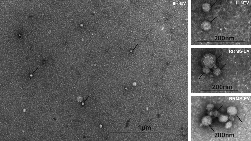
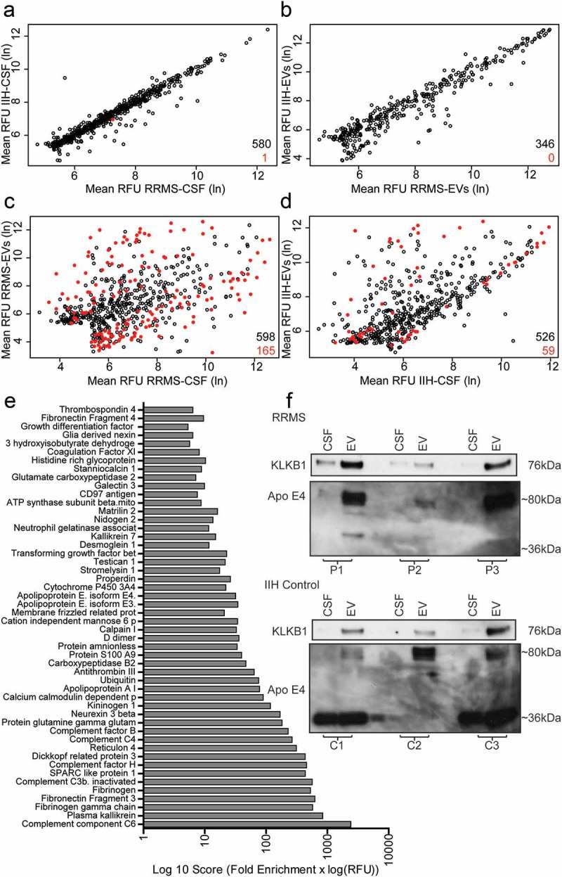
The discovery of disease biomarkers, along with the use of "liquid biopsies" as a minimally invasive source of biomarkers, continues to be of great interest. In inflammatory diseases of the central nervous system (CNS), cerebrospinal fluid (CSF) is the most obvious biofluid source. Extracellular vesicles (EVs) are also present in CSF and are thought to be potential "biomarker treasure chests". However, isolating these CSF-derived EVs remains challenging. This small-scale pilot study developed and tested a protocol to enrich for CSF-EVs, both in relapsing remitting multiple sclerosis (RRMS) CSF and controls. These were subsequently compared, using an aptamer based proteomics array, SOMAscan™. EVs were enriched from RRMS patient ( = 4) and non-demyelinating control (idiopathic intracranial hypertension (IIH) ( = 3)) CSF using precipitation and mini size-exclusion chromatography (SEC). EV-enriched fractions were selected using pre-defined EV characteristics, including increased levels of tetraspanins. EVs and paired CSF were analysed by SOMAscan™, providing relative abundance data for 1128 proteins. CSF-EVs were characterised, revealing exosome-like features: rich in tetraspanins CD9 and CD81, size ~100 nm, and exosome-like morphology by TEM. Sufficient quantities of, SOMAscan™ compatible, EV material was obtained from 5 ml CSF for proteomics analysis. Overall, 348 and 580 proteins were identified in CSF-EVs and CSF, respectively, of which 50 were found to be significantly (-test) and exclusively enriched in RRMS CSF-EVs. Selected proteins, Plasma kallikrein and Apolipoprotein-E4, were further validated by western blot and appeared increased in CSF-EVs compared to CSF. Functional enrichment analysis of the 50 enriched proteins revealed strong associations with biological processes relating to MS pathology and also extracellular regions, consistent with EV enrichment. This pilot study demonstrates practicality for EV enrichment in CSF derived from patients with MS and controls, allowing detailed analysis of protein profiles that may offer opportunities to identify novel biomarkers and therapeutic approaches in CNS inflammatory diseases.
疾病生物标志物的发现,以及将“液体活检”作为生物标志物的微创来源的应用,仍然备受关注。在中枢神经系统(CNS)的炎症性疾病中,脑脊液(CSF)是最明显的生物流体来源。细胞外囊泡(EVs)也存在于脑脊液中,被认为是潜在的“生物标志物宝库”。然而,分离这些源自脑脊液的细胞外囊泡仍然具有挑战性。这项小规模的试点研究开发并测试了一种方案,用于富集复发缓解型多发性硬化症(RRMS)脑脊液和对照中的脑脊液来源的细胞外囊泡。随后,使用基于适配体的蛋白质组学阵列SOMAscan™对这些样本进行了比较。通过沉淀和微型尺寸排阻色谱法(SEC)从RRMS患者(n = 4)和非脱髓鞘对照(特发性颅内高压(IIH)(n = 3))的脑脊液中富集细胞外囊泡。使用预先定义的细胞外囊泡特征(包括四跨膜蛋白水平升高)选择富含细胞外囊泡的组分。通过SOMAscan™分析细胞外囊泡和配对的脑脊液,提供1128种蛋白质的相对丰度数据。对脑脊液来源的细胞外囊泡进行了表征,揭示了类似外泌体的特征:富含四跨膜蛋白CD9和CD81,大小约为100nm,通过透射电子显微镜观察具有类似外泌体的形态。从5ml脑脊液中获得了足够量的、与SOMAscan™兼容的细胞外囊泡材料用于蛋白质组学分析。总体而言,在脑脊液来源的细胞外囊泡和脑脊液中分别鉴定出348种和580种蛋白质,其中50种被发现显著(t检验)且仅在RRMS脑脊液来源的细胞外囊泡中富集。通过蛋白质免疫印迹进一步验证了选定的蛋白质血浆激肽释放酶和载脂蛋白E4,与脑脊液相比,它们在脑脊液来源的细胞外囊泡中含量增加。对这50种富集蛋白质的功能富集分析揭示了与MS病理相关的生物学过程以及细胞外区域的强关联,这与细胞外囊泡的富集一致。这项试点研究证明了在MS患者和对照的脑脊液中富集细胞外囊泡的实用性,允许对蛋白质谱进行详细分析,这可能为识别中枢神经系统炎症性疾病中的新型生物标志物和治疗方法提供机会。