Kunzke Thomas, Balluff Benjamin, Feuchtinger Annette, Buck Achim, Langer Rupert, Luber Birgit, Lordick Florian, Zitzelsberger Horst, Aichler Michaela, Walch Axel
Research Unit Analytical Pathology, Helmholtz Zentrum München, OberschleiΔheim, Germany.
Maastricht MultiModal Molecular Imaging Institute (M4I), Maastricht University, Maastricht, The Netherlands.
Oncotarget. 2017 Jul 10;8(40):68012-68025. doi: 10.18632/oncotarget.19137. eCollection 2017 Sep 15.
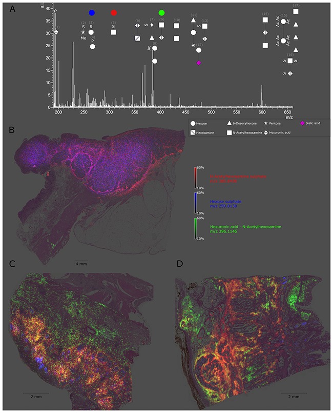
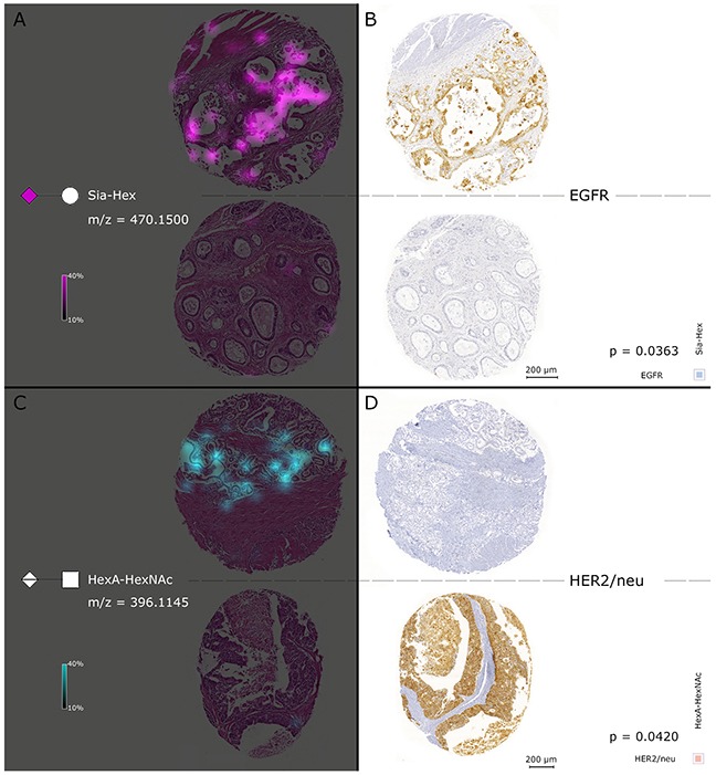
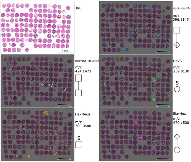
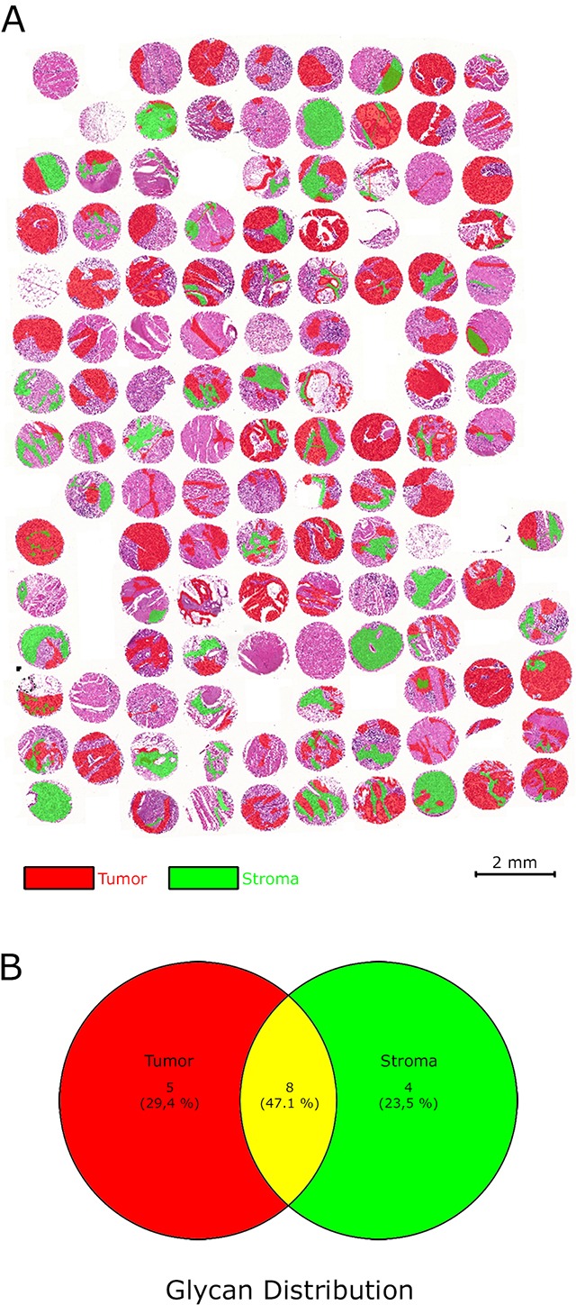
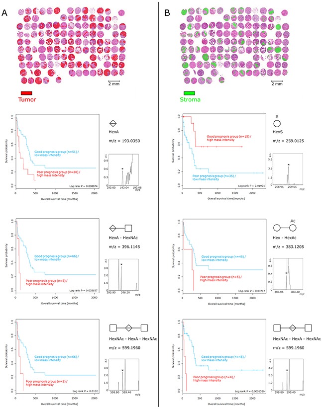
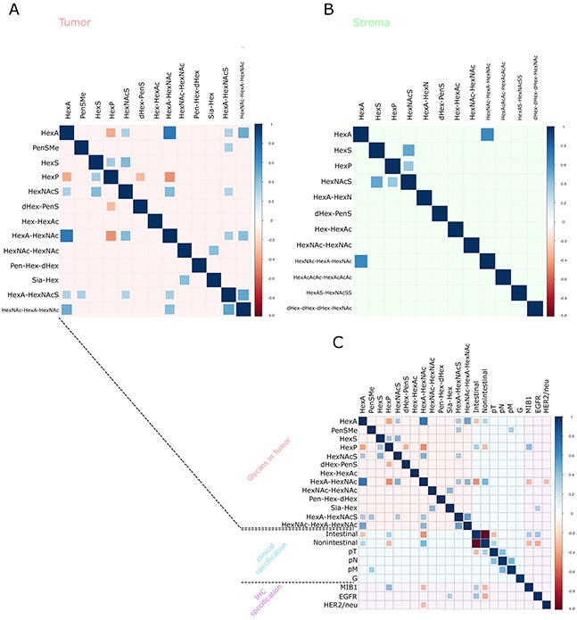
Glycosylation in cancer is a highly dynamic process that has a significant impact on tumor biology. Further, the attachment of aberrant glycan forms is already considered a hallmark of the disease state. Mass spectrometry has become a prominent approach to analyzing glycoconjugates. Specifically, matrix-assisted laser desorption/ionisation -mass spectrometric imaging (MALDI-MSI) is a powerful technique that combines mass spectrometry with histology and enables the spatially resolved and label-free detection of glycans. The most common approach to the analysis of glycans is the use of mass spectrometry adjunct to PNGase F digestion and other chemical reactions. In the current study, we perform the analysis of formalin-fixed, paraffin-embedded (FFPE) tissues for natively occurring bioactive glycan fragments without prior digestion or chemical reactions using MALDI-FT-ICR-MSI. We examined 106 primary resected gastric cancer patient tissues in a tissue microarray and correlated native-occurring fragments with clinical endpoints, therapeutic targets such as epidermal growth factor receptor (EGFR) and HER2/neu expressions and the proliferation marker MIB1. The detection of a glycosaminoglycan fragment in tumor stroma regions was determined to be an independent prognostic factor for gastric cancer patients. Native glycan fragments were significantly linked to the expression of EGFR, HER2/neu and MIB1. In conclusion, we are the first to report the detection of native-occurring bioactive glycan fragments in FFPE tissues that influence patient outcomes. These findings highlight the significance of glycan fragments in gastric cancer tumor biology and patient outcome.
癌症中的糖基化是一个高度动态的过程,对肿瘤生物学有重大影响。此外,异常聚糖形式的附着已被视为疾病状态的一个标志。质谱已成为分析糖缀合物的一种重要方法。具体而言,基质辅助激光解吸/电离质谱成像(MALDI-MSI)是一种强大的技术,它将质谱与组织学相结合,能够对聚糖进行空间分辨且无需标记的检测。分析聚糖最常用的方法是将质谱与PNGase F消化及其他化学反应相结合。在本研究中,我们使用MALDI-FT-ICR-MSI对福尔马林固定、石蜡包埋(FFPE)组织中天然存在的生物活性聚糖片段进行分析,无需事先消化或化学反应。我们在组织芯片中检测了106例原发性切除的胃癌患者组织,并将天然存在的片段与临床终点、治疗靶点(如表皮生长因子受体(EGFR)和HER2/neu表达)以及增殖标志物MIB1进行关联。肿瘤基质区域中一种糖胺聚糖片段的检测被确定为胃癌患者的独立预后因素。天然聚糖片段与EGFR、HER2/neu和MIB1的表达显著相关。总之,我们首次报道了在FFPE组织中检测到影响患者预后的天然存在的生物活性聚糖片段。这些发现突出了聚糖片段在胃癌肿瘤生物学和患者预后中的重要性。