• 文献检索
  • 文档翻译
  • 深度研究
  • 学术资讯
  • Suppr Zotero 插件Zotero 插件
  • 邀请有礼
  • 套餐&价格
  • 历史记录
应用&插件
Suppr Zotero 插件Zotero 插件浏览器插件Mac 客户端Windows 客户端微信小程序
定价
高级版会员购买积分包购买API积分包
服务
文献检索文档翻译深度研究API 文档MCP 服务
关于我们
关于 Suppr公司介绍联系我们用户协议隐私条款
关注我们

Suppr 超能文献

核心技术专利:CN118964589B侵权必究
粤ICP备2023148730 号-1Suppr @ 2026

文献检索

告别复杂PubMed语法,用中文像聊天一样搜索,搜遍4000万医学文献。AI智能推荐,让科研检索更轻松。

立即免费搜索

文件翻译

保留排版,准确专业,支持PDF/Word/PPT等文件格式,支持 12+语言互译。

免费翻译文档

深度研究

AI帮你快速写综述,25分钟生成高质量综述,智能提取关键信息,辅助科研写作。

立即免费体验

使用锥形束计算机断层扫描分析上颌切牙在牙槽骨中的牙根位置。

Analysis of the root position of the maxillary incisors in the alveolar bone using cone-beam computed tomography.

作者信息

Jung Yun-Hoa, Cho Bong-Hae, Hwang Jae Joon

机构信息

Department of Oral and Maxillofacial Radiology, School of Dentistry, Pusan National University, Yangsan, Korea.

Department of Oral and Maxillofacial Radiology, Yonsei University College of Dentistry, Seoul, Korea.

出版信息

Imaging Sci Dent. 2017 Sep;47(3):181-187. doi: 10.5624/isd.2017.47.3.181. Epub 2017 Sep 21.

DOI:10.5624/isd.2017.47.3.181
PMID:28989901
原文链接:https://pmc.ncbi.nlm.nih.gov/articles/PMC5620463/
Abstract

PURPOSE

The purpose of this study was to measure the buccal bone thickness and angulation of the maxillary incisors and to analyze the correlation between these parameters and the root position in the alveolar bone using cone-beam computed tomography (CBCT).

MATERIALS AND METHODS

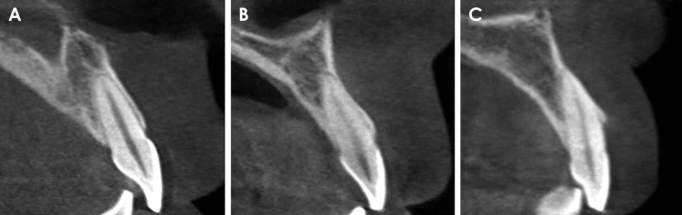
CBCT images of 398 maxillary central and lateral incisors from 199 patients were retrospectively reviewed. The root position in the alveolar bone was classified as buccal, middle, or palatal, and the buccal type was further classified into subtypes I, II, and III. In addition, the buccolingual inclination of the tooth and buccal bone thickness were evaluated.

RESULTS

A majority of the maxillary incisors were positioned more buccally within the alveolar bone, and only 2 lateral incisors (0.5%) were positioned more palatally. The angulation of buccal subtype III was the greatest and that of the middle type was the lowest. Most of the maxillary incisors exhibited a thin facial bone wall, and the lateral incisors had a significantly thinner buccal bone than the central incisors. The buccal bone of buccal subtypes II and III was significantly thinner than that of buccal subtype I.

CONCLUSION

A majority of the maxillary incisor roots were positioned close to the buccal cortical plate and had a thin buccal bone wall. Significant relationships were observed between the root position in the alveolar bone, the angulation of the tooth in the alveolar bone, and buccal bone thickness. CBCT analyses of the buccal bone and sagittal root position are recommended for the selection of the appropriate treatment approach.

摘要

目的

本研究旨在使用锥形束计算机断层扫描(CBCT)测量上颌切牙的颊侧骨厚度和角度,并分析这些参数与牙槽骨中牙根位置之间的相关性。

材料与方法

回顾性分析了199例患者的398颗上颌中切牙和侧切牙的CBCT图像。牙槽骨中的牙根位置分为颊侧、中间或腭侧,颊侧类型进一步分为I、II和III亚型。此外,评估了牙齿的颊舌倾斜度和颊侧骨厚度。

结果

大多数上颌切牙在牙槽骨内的位置更靠近颊侧,只有2颗侧切牙(0.5%)的位置更靠近腭侧。颊侧III型的角度最大,中间型的角度最小。大多数上颌切牙表现出较薄的面部骨壁,侧切牙的颊侧骨明显比中切牙薄。颊侧II型和III型的颊侧骨明显比颊侧I型薄。

结论

大多数上颌切牙根靠近颊侧皮质板,颊侧骨壁较薄。牙槽骨中的牙根位置、牙槽骨中牙齿的角度和颊侧骨厚度之间存在显著关系。建议对颊侧骨和矢状牙根位置进行CBCT分析,以选择合适的治疗方法。

https://cdn.ncbi.nlm.nih.gov/pmc/blobs/c9da/5620463/d04ffefdf291/isd-47-181-g004.jpg
https://cdn.ncbi.nlm.nih.gov/pmc/blobs/c9da/5620463/d2804ec41a2f/isd-47-181-g001.jpg
https://cdn.ncbi.nlm.nih.gov/pmc/blobs/c9da/5620463/97783e9a008b/isd-47-181-g002.jpg
https://cdn.ncbi.nlm.nih.gov/pmc/blobs/c9da/5620463/5787e1436696/isd-47-181-g003.jpg
https://cdn.ncbi.nlm.nih.gov/pmc/blobs/c9da/5620463/d04ffefdf291/isd-47-181-g004.jpg

相似文献

1
Analysis of the root position of the maxillary incisors in the alveolar bone using cone-beam computed tomography.使用锥形束计算机断层扫描分析上颌切牙在牙槽骨中的牙根位置。
Imaging Sci Dent. 2017 Sep;47(3):181-187. doi: 10.5624/isd.2017.47.3.181. Epub 2017 Sep 21.
2
Assessment of Maxillary Incisors' Angulation and Position in Different Types of Malocclusions Using Cone-Beam Computed Tomography.使用锥形束计算机断层扫描评估不同类型错牙合畸形中上颌切牙的角度和位置
Contemp Clin Dent. 2021 Oct-Dec;12(4):401-407. doi: 10.4103/ccd.ccd_743_20. Epub 2021 Dec 21.
3
[Classification of the anatomical structures of maxillary central incisor root by cone-beam CT].[锥形束CT对上颌中切牙根解剖结构的分类]
Zhonghua Kou Qiang Yi Xue Za Zhi. 2017 Nov 9;52(11):656-660. doi: 10.3760/cma.j.issn.1002-0098.2017.11.002.
4
[Investigation of sagittal root position in relation to the anterior maxillary alveolar bone: a cone-beam CT study in 300 cases with normal occlusion].[矢状面牙根位置与上颌前牙区牙槽骨关系的研究:300例正常咬合患者的锥形束CT研究]
Zhonghua Kou Qiang Yi Xue Za Zhi. 2017 Oct 9;52(10):631-636. doi: 10.3760/cma.j.issn.1002-0098.2017.10.010.
5
Analysis of the root position and angulation of maxillary premolars in alveolar bone using cone-beam computed tomography.使用锥形束计算机断层扫描分析上颌前磨牙在牙槽骨中的牙根位置和角度。
Imaging Sci Dent. 2022 Dec;52(4):365-373. doi: 10.5624/isd.20220710. Epub 2022 Sep 7.
6
Clinical assessment of the palatal alveolar bone thickness and its correlation with the buccolingual angulation of maxillary incisors for immediate implant placement.即刻种植时评估腭侧牙槽骨厚度及其与上颌切牙颊舌向倾斜角度的临床相关性。
Clin Implant Dent Relat Res. 2019 Oct;21(5):1080-1086. doi: 10.1111/cid.12835. Epub 2019 Aug 19.
7
Comparison of alveolar bone width and sagittal tooth angulation of maxillary central incisors in Class I and Class III canine relationships: a retrospective study using CBCT.使用 CBCT 对 I 类和 III 类犬关系中上颌中切牙牙槽骨宽度和矢状牙倾斜角的比较:回顾性研究。
BMC Oral Health. 2022 Jul 22;22(1):303. doi: 10.1186/s12903-022-02331-x.
8
Influence of alveolar bone thickness and bucco-palatal inclination on root resorption of lateral incisors in unilateral maxillary impacted canines: a retrospective observational study.单侧上颌埋伏尖牙中牙槽骨厚度和颊腭向倾斜度对侧切牙根吸收的影响:一项回顾性观察研究。
BMC Oral Health. 2024 Mar 2;24(1):301. doi: 10.1186/s12903-024-04076-1.
9
Classification of the Root Position of the Maxillary Central Incisors and its Clinical Significance in Immediate Implant Placement.上颌中切牙根位置分类及其在即刻种植中的临床意义
Implant Dent. 2016 Aug;25(4):520-4. doi: 10.1097/ID.0000000000000438.
10
Labial alveolar bone thickness and its correlation with buccolingual maxillary incisors angulation: a CBCT based study.基于 CBCT 的研究:唇牙槽骨厚度及其与上颌切牙颊舌向倾斜角度的相关性。
Eur Rev Med Pharmacol Sci. 2022 Jul;26(13):4625-4633. doi: 10.26355/eurrev_202207_29184.

引用本文的文献

1
Analysis of the Sagittal Root Position of the Maxillary and Mandibular Anterior Teeth in the Alveolar Bone Using Cone-Beam Computed Tomography.使用锥形束计算机断层扫描分析上颌和下颌前牙在牙槽骨中的矢状根位置。
Diagnostics (Basel). 2024 Dec 6;14(23):2756. doi: 10.3390/diagnostics14232756.
2
Evaluation of the anterior dentoalveolar relationship in skeletal Class III malocclusion patients with different vertical facial patterns using cone-beam computed tomography.应用锥形束 CT 评价不同垂直面型的骨性 III 类错颌患者的前牙牙槽骨关系。
Angle Orthod. 2024 Mar 1;94(2):187-193. doi: 10.2319/081723-563.1.
3
Evaluation of alveolar bone morphology of incisors with different sagittal skeletal facial types by cone beam computed tomography: A retrospective study.

本文引用的文献

1
Classification of the Root Position of the Maxillary Central Incisors and its Clinical Significance in Immediate Implant Placement.上颌中切牙根位置分类及其在即刻种植中的临床意义
Implant Dent. 2016 Aug;25(4):520-4. doi: 10.1097/ID.0000000000000438.
2
Buccal Bone Thickness Overlying Maxillary Anterior Teeth: A Clinical and Radiographic Prospective Human Study.上颌前牙区颊侧骨厚度:一项临床与影像学前瞻性人体研究。
Implant Dent. 2016 Aug;25(4):525-31. doi: 10.1097/ID.0000000000000427.
3
Preoperative assessment of labial bone perforation for virtual immediate implant surgery in the maxillary esthetic zone: A computer simulation study.
锥形束计算机断层扫描评估不同矢状骨骼面型切牙的牙槽骨形态:一项回顾性研究。
Heliyon. 2023 Apr 10;9(4):e15369. doi: 10.1016/j.heliyon.2023.e15369. eCollection 2023 Apr.
4
Efficacy of upper-incisor torque control with clear aligners: a retrospective study using cone-beam computed tomography.使用透明牙套控制上前牙转矩的疗效:一项基于锥形束 CT 的回顾性研究。
Clin Oral Investig. 2023 Jul;27(7):3863-3873. doi: 10.1007/s00784-023-05006-1. Epub 2023 Apr 10.
5
Are morphological and morphometric characteristics of maxillary anterior region and nasopalatine canal related to each other?上颌前部区域和鼻腭管的形态学及形态测量学特征彼此相关吗?
Oral Radiol. 2023 Apr;39(2):372-385. doi: 10.1007/s11282-022-00647-6. Epub 2022 Aug 8.
6
Tomographic sagittal root position in relation to maxillary anterior bone housing in a Brazilian population.巴西人群中根尖片矢状位牙根相对于上颌前部骨容纳结构的位置
Imaging Sci Dent. 2022 Mar;52(1):75-82. doi: 10.5624/isd.20210226. Epub 2022 Feb 11.
7
Assessment of Maxillary Incisors' Angulation and Position in Different Types of Malocclusions Using Cone-Beam Computed Tomography.使用锥形束计算机断层扫描评估不同类型错牙合畸形中上颌切牙的角度和位置
Contemp Clin Dent. 2021 Oct-Dec;12(4):401-407. doi: 10.4103/ccd.ccd_743_20. Epub 2021 Dec 21.
8
Facial alveolar bone thickness and modifying factors of anterior maxillary teeth: a systematic review and meta-analysis of cone-beam computed tomography studies.面部牙槽骨厚度和上前牙的影响因素:锥形束 CT 研究的系统评价和荟萃分析。
BMC Oral Health. 2021 Mar 22;21(1):143. doi: 10.1186/s12903-021-01495-2.
9
Influence of the anterior arch shape and root position on root angulation in the maxillary esthetic area.上颌美学区域前牙弓形态及牙根位置对牙根角度的影响。
Imaging Sci Dent. 2019 Jun;49(2):123-130. doi: 10.5624/isd.2019.49.2.123. Epub 2019 Jun 24.
10
Correlation of anterior overbite with root position and buccal bone thickness of maxillary anterior teeth: a CBCT study.上颌前牙覆合与牙根位置及颊侧骨厚度的相关性:一项CBCT研究
Surg Radiol Anat. 2019 Aug;41(8):935-942. doi: 10.1007/s00276-019-02257-0. Epub 2019 May 25.
上颌美学区虚拟即刻种植手术唇侧骨穿孔的术前评估:一项计算机模拟研究。
J Am Dent Assoc. 2015 Nov;146(11):808-19. doi: 10.1016/j.adaj.2015.04.007.
4
Preoperative implant planning considering alveolar bone grafting needs and complication prediction using panoramic versus CBCT images.考虑牙槽骨移植需求的术前种植体规划以及使用全景片与锥形束计算机断层扫描(CBCT)图像进行并发症预测。
Imaging Sci Dent. 2014 Sep;44(3):213-20. doi: 10.5624/isd.2014.44.3.213. Epub 2014 Sep 17.
5
Analysis of facial bone wall dimensions and sagittal root position in the maxillary esthetic zone: a retrospective study using cone beam computed tomography.上颌美学区面部骨壁尺寸及牙根矢状位置分析:一项使用锥形束计算机断层扫描的回顾性研究
Int J Oral Maxillofac Implants. 2014 Sep-Oct;29(5):1123-9. doi: 10.11607/jomi.3348.
6
Determination of implant position for immediate implant placement in maxillary central incisors using palatal soft tissue landmarks.利用腭侧软组织解剖标志确定上颌中切牙即刻种植的种植体位置。
Int J Oral Maxillofac Implants. 2014 May-Jun;29(3):627-33. doi: 10.11607/jomi.2907.
7
Incidence of implant buccal plate fenestration in the esthetic zone: a cone beam computed tomography study.美学区种植体颊侧穿龈缺损的发生率:锥形束 CT 研究。
Int J Oral Maxillofac Implants. 2014 Jan-Feb;29(1):171-7. doi: 10.11607/jomi.3397.
8
Analysis of the dimensions of the labial bone wall in the anterior maxilla: a cone-beam computed tomography study.上颌前部唇侧骨壁尺寸分析:一项锥形束计算机断层扫描研究。
Clin Oral Implants Res. 2015 Apr;26(4):e57-e61. doi: 10.1111/clr.12332. Epub 2014 Jan 23.
9
Facial alveolar bone wall width - a cone-beam computed tomography study in Asians.亚洲人颊牙槽骨壁宽度的锥形束 CT 研究。
Clin Oral Implants Res. 2014 Feb;25(2):194-206. doi: 10.1111/clr.12096. Epub 2013 Jan 7.
10
Evaluation of buccal alveolar bone dimension of maxillary anterior and premolar teeth: a cone beam computed tomography investigation.上颌前牙和前磨牙颊牙槽骨尺寸的评估:锥形束 CT 研究。
Int J Oral Maxillofac Implants. 2012 Nov-Dec;27(6):1514-9.