Liu Jianhua, Sui Hua, Zhao Jianlin, Wang Yan
Department of Cardiology, Xinxiang Central HospitalXinxiang, Henan, China.
Department of Endocrinology, Xinxiang Central HospitalXinxiang, Henan, China.
Front Physiol. 2017 Sep 25;8:611. doi: 10.3389/fphys.2017.00611. eCollection 2017.
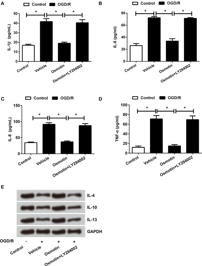
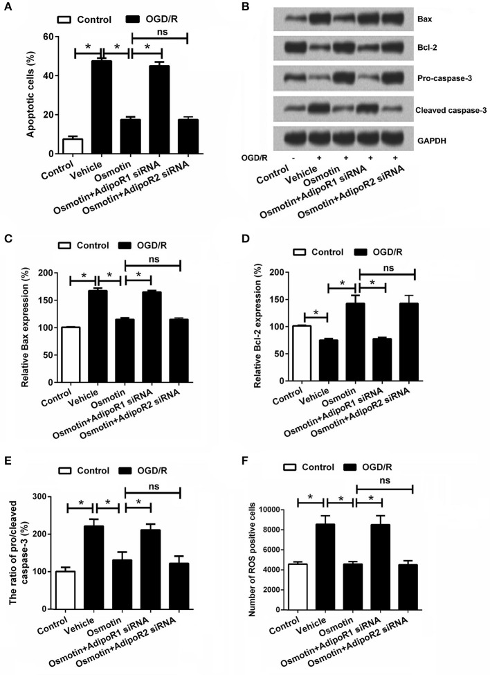
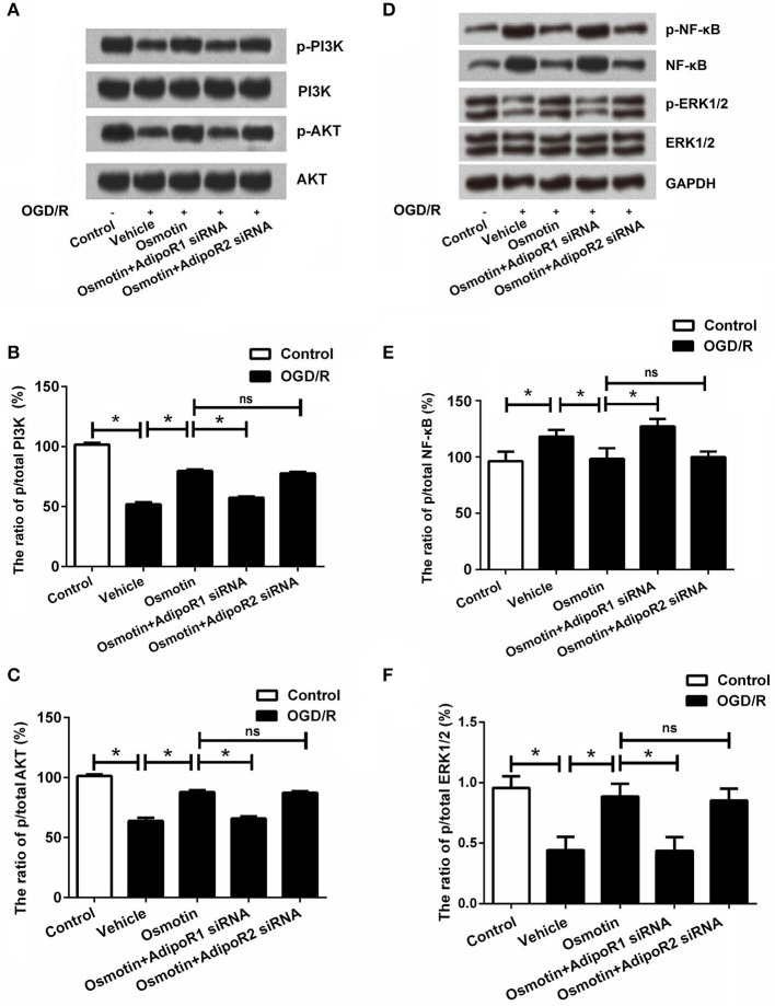
This study aimed to investigate the effect of osmotin on myocardial ischemia/reperfusion (I/R), as well as the underlying mechanisms. I/R injury model was established on rat cardiac myoblast H9c2 cells by oxygen and glucose deprivation followed by reperfusion (OGD/R). Cells were administrated with osmotin, and transfected with small interfering RNAs (siRNAs) which specifically target adiponectin receptor 1 or 2 (AdipoR1/2). Besides, the cells were incubated with or without LY294002 as inhibitor of phosphatidylinositol 3-kinase (PI3K) under OGD/R condition. Cell viability, apoptosis, expressions of apoptosis-related proteins and inflammatory factors were analyzed. The results showed that osmotin significantly increased H9c2 cells viability compared with the cells treated with vehicle ( < 0.05), and decreased H9c2 cells apoptosis by regulating expressions of apoptosis-related proteins. Moreover, we observed that osmotin statistically reduced the release of proinflammatory factors and increased the release of anti-inflammatory factors in H9c2 cells ( < 0.05). However, these effects were markedly reversed by AdipoR1 silence but not AdipoR2. Furthermore, osmotin dramatically upregulated the phosphorylation levels of PI3K, AKT, ERK, and downregulated the phosphorylation level of NF-κB ( < 0.05). While administration of LY294002 reduced cell viability, increased cell apoptosis, and aggravated inflammatory response ( < 0.05). Our results suggested that the protective effect of osmotin on the simulated OGD/R injured H9c2 cells might be associated with AdipoR1/PI3K/AKT signaling pathway.
本研究旨在探讨渗透蛋白对心肌缺血/再灌注(I/R)的影响及其潜在机制。通过氧糖剥夺复氧(OGD/R)建立大鼠心肌成肌细胞H9c2细胞的I/R损伤模型。细胞用渗透蛋白处理,并用特异性靶向脂联素受体1或2(AdipoR1/2)的小干扰RNA(siRNA)转染。此外,在OGD/R条件下,细胞在有或没有作为磷脂酰肌醇3激酶(PI3K)抑制剂的LY294002的情况下孵育。分析细胞活力、凋亡、凋亡相关蛋白和炎性因子的表达。结果表明,与用溶媒处理的细胞相比,渗透蛋白显著提高了H9c2细胞的活力(P<0.05),并通过调节凋亡相关蛋白的表达降低了H9c2细胞的凋亡。此外,我们观察到渗透蛋白在统计学上降低了H9c2细胞中促炎因子的释放并增加了抗炎因子的释放(P<0.05)。然而,这些作用被AdipoR1沉默显著逆转,但AdipoR2沉默则不然。此外,渗透蛋白显著上调了PI3K、AKT、ERK的磷酸化水平,并下调了NF-κB的磷酸化水平(P<0.05)。而给予LY294002降低了细胞活力,增加了细胞凋亡,并加重了炎症反应(P<0.05)。我们的结果表明,渗透蛋白对模拟OGD/R损伤的H9c2细胞的保护作用可能与AdipoR1/PI3K/AKT信号通路有关。