El-Gerby Khaled M, El-Anwar Mohammad Waheed
Radiodiagnosis Department, Faculty of Medicine, Zagazig University, Zagazig, Egypt.
Department of Otorhinolaryngology Head and Neck Surgery, Faculty of Medicine, Zagazig University, Zagazig, Egypt.
Int Arch Otorhinolaryngol. 2017 Oct;21(4):358-365. doi: 10.1055/s-0036-1597323. Epub 2017 Jan 4.
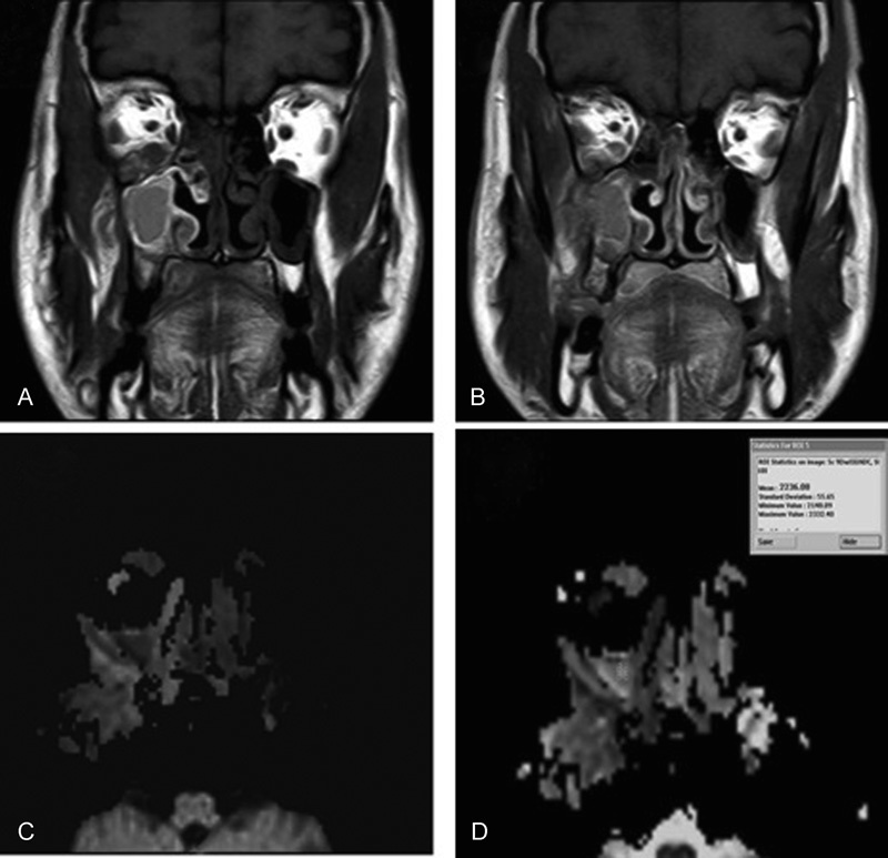
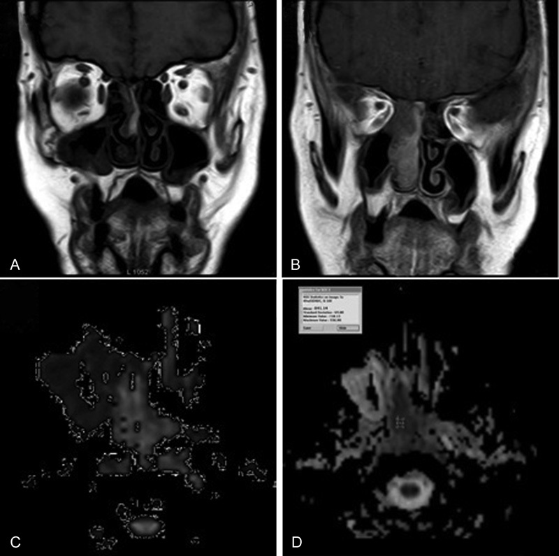
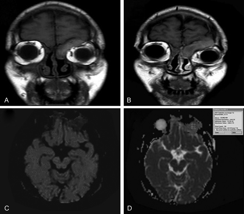
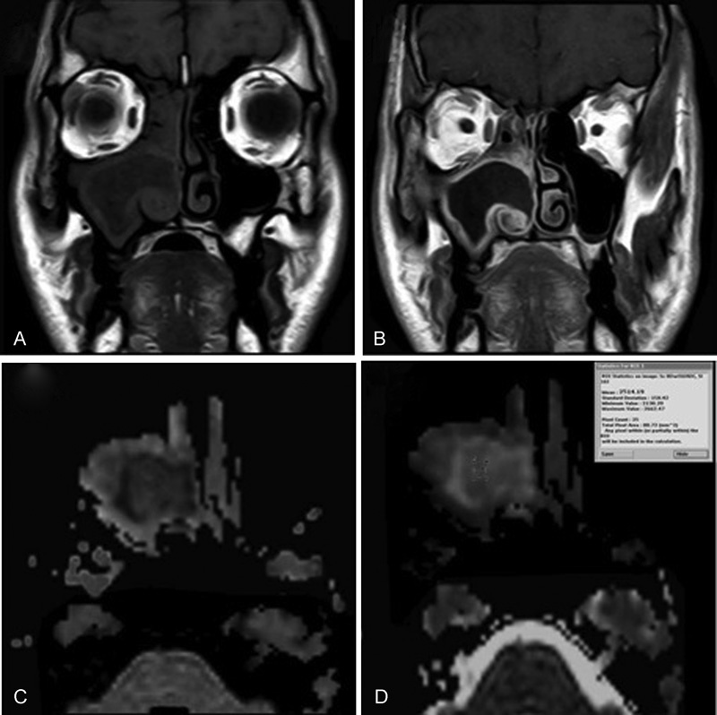
Appearance of nasal masses on routine CT and MRI are not pathognomonic. We utilized the apparent diffusion coefficient (ADC) value obtained from diffusion weighted image (DWI) to detect the differences in the microstructures of tumor and non-tumor tissues. The objective of our study was to evaluate the diagnostic role of DWI and ADC values in differentiating between malignant and benign sinonasal lesions and its correlation with histopathological results as the reference standard. Patients with nasal and / or paranasal mass underwent CT, MRI, and DWI before any surgical intervention. We used diagnostic sinonasal endoscopy and biopsy to confirm the diagnosis after MRI. When we used ADC value of (1.2 × 10⁻³ mm2/s) as a cut-off value for differentiating benign from malignant sinonasal lesions, we achieved 90% accuracy, 100% sensitivity, 88.4% specificity, 77.8% positive predictive value, and 100% negative predictive value. At this cut-off, benign lesions show statistically significant higher ADC value than malignant tumors. DW MRI and ADC value calculation are promising quantitative methods helping to differentiate between malignant and benign sinonasal lesions. Thus, they are effective methods compared with other conventional methods with short imaging time thus it is recommended to be incorporated into routine evaluations.
常规CT和MRI上鼻肿块的表现并无特异性。我们利用从扩散加权成像(DWI)获得的表观扩散系数(ADC)值来检测肿瘤组织和非肿瘤组织微观结构的差异。
我们研究的目的是评估DWI和ADC值在鉴别鼻窦良恶性病变中的诊断作用,以及其与作为参考标准的组织病理学结果的相关性。
患有鼻和/或鼻窦肿块的患者在任何手术干预前均接受了CT、MRI和DWI检查。我们在MRI检查后使用诊断性鼻内镜检查和活检来确诊。
当我们将(1.2×10⁻³ mm2/s)的ADC值作为区分鼻窦良恶性病变的临界值时,准确率达到90%,敏感性为100%,特异性为88.4%,阳性预测值为77.8%,阴性预测值为100%。在此临界值下,良性病变的ADC值在统计学上显著高于恶性肿瘤。
DW MRI和ADC值计算是有助于鉴别鼻窦良恶性病变的有前景的定量方法。因此,与其他传统方法相比,它们是有效的方法,成像时间短,因此建议纳入常规评估。