Wang Chih-Ping, Lai Chien-Hsiung, Huang Evelyn Jou-Chen, Wu Pei-Lun, Chen Ching-Lung, Chen Chau-Yin, King Yin-Chi, Wu Pei-Chen, Kuo Chien-Neng
Department of Ophthalmology, Chang Gung Memorial Hospital, Chiayi, Taiwan.
College of Medicine, Chang Gung University, Tao-Yuan, Taiwan.
Taiwan J Ophthalmol. 2015 Oct-Dec;5(4):169-176. doi: 10.1016/j.tjo.2015.10.002. Epub 2015 Nov 30.
To compare axial length (AL) and subfoveal choroidal thickness (SFCT) between individuals with age-related macular degeneration (AMD) and controls with no lesions.
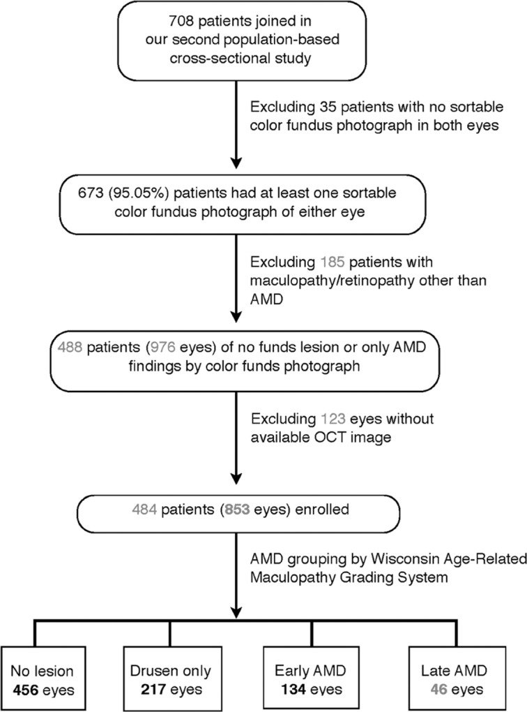
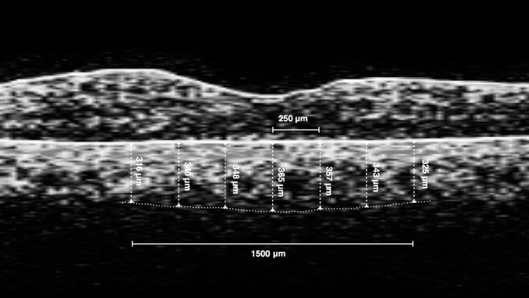
This was a case-control study. In total, 853 eyes of 484 patients (>65 years), including 397 eyes at various AMD stages and 456 eyes with no fundus lesions (controls) were recruited. Using color fundus photography, eyes were grouped according to AMD degree. AL was automatically measured using IOL Master and SFCT was manually measured by two independent observers. The associations among age, AL, SFCT, and each AMD grade were analyzed.
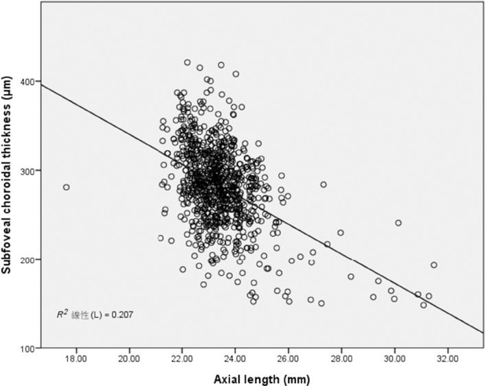
Out of 853 eyes, 456 had no lesions, 217 contained drusen only, 134 had early AMD, and 46 had late AMD. The eyes with late AMD were older ( = 0.007) and had longer AL ( ≥ 0.001) and thinner SFCT ( < 0.001) compared with groups of no fundus lesions, drusen only, and early AMD. SFCT in eyes with late AMD decreased by 19.20 μm ( = 0.049), 24.78 μm ( = 0.029), and 15.56 μm ( = 0.162) compared with groups of no fundus lesions, drusen only, and early AMD, respectively. SFCT decreased by 14.18 μm/mm increase in AL ( < 0.001). The odds ratio (OR) for late AMD by longer AL (≥25 mm) and thinner SFCT (<240 μm) was 4.54 (χ = 9.36; = 0.002) and 4.86 (χ = 17.62; < 0.001), respectively, and was 9.57 (χ = 18.07; < 0.001) when both AL ≥ 25 ≥m and SFCT < 240 μm.
Eyes with late AMD have distinct reduced SFCT and elongated AL. Eyes with thinner SFCT and longer AL showed high ORs for late AMD and even higher ORs when both factors were simultaneously present. These findings illustrate the crucial pathophysiological role of these two important ocular fac tors and arouse our attention to patients with both characteristics, especially in Asian countries where the prevalence of myopia are disturbingly high.
比较年龄相关性黄斑变性(AMD)患者与无病变对照者的眼轴长度(AL)和黄斑中心凹下脉络膜厚度(SFCT)。
这是一项病例对照研究。共纳入484例年龄>65岁患者的853只眼,其中包括处于不同AMD阶段的397只眼和456只无眼底病变的眼(对照)。使用彩色眼底照相术,根据AMD程度对眼睛进行分组。使用IOL Master自动测量AL,由两名独立观察者手动测量SFCT。分析年龄、AL、SFCT与各AMD分级之间的关联。
在853只眼中,456只无病变,217只仅有玻璃膜疣,134只患有早期AMD,46只患有晚期AMD。与无眼底病变、仅有玻璃膜疣和早期AMD组相比,晚期AMD患者的年龄更大(P = 0.007),AL更长(P≥0.001),SFCT更薄(P<0.001)。与无眼底病变、仅有玻璃膜疣和早期AMD组相比,晚期AMD患者的SFCT分别降低了19.20μm(P = 0.049)、24.78μm(P = 0.029)和15.56μm(P = 0.162)。AL每增加1mm,SFCT降低14.18μm(P<0.001)。AL较长(≥25mm)和SFCT较薄(<240μm)的晚期AMD的比值比(OR)分别为4.54(χ² = 9.36;P = 0.002)和4.86(χ² = 17.62;P<0.001),当AL≥25mm且SFCT<240μm时,OR为9.57(χ² = 18.07;P<0.001)。
晚期AMD患者的SFCT明显降低,AL延长。SFCT较薄且AL较长的眼发生晚期AMD的OR较高,当这两个因素同时存在时OR更高。这些发现说明了这两个重要眼部因素的关键病理生理作用,并引起我们对具有这两种特征患者的关注,尤其是在近视患病率高得惊人的亚洲国家。