Department of Medical Physics and Biomedical Engineering, University College London, Gower Street, London, WC1E 6BT, UK.
Royal London Hospital, Barts Health NHS Trust, Whitechapel Road, London, E1 1BB, UK.
Sci Rep. 2017 Oct 11;7(1):12998. doi: 10.1038/s41598-017-13399-9.
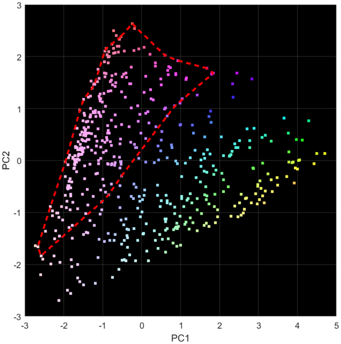
This pilot study examines the correlation of X-ray diffraction (XRD) measurements with the histopathological analysis of breast tissue. Eight breast cancer samples were investigated. Each sample contained a mixture of normal and cancerous tissues. In total, 522 separate XRD measurements were made at different locations across the samples (8 in total). The resulting XRD spectra were subjected to principal component analysis (PCA) in order to determine if there were any distinguishing features that could be used to identify different tissue components. 99.0% of the variation between the spectra were described by the first two principal components (PC). Comparing the location of points in PC space with the classification determined by histopathology indicated correlation between the shape/magnitude of the XRD spectra and the tissue type. These results are encouraging and suggest that XRD could be used for the intraoperative or postoperative classification of bulk tissue samples.
本初步研究考察了 X 射线衍射(XRD)测量与乳腺组织的组织病理学分析之间的相关性。研究了 8 个乳腺癌样本。每个样本都包含正常组织和癌变组织的混合物。总共在样本的不同位置进行了 522 次单独的 XRD 测量(共 8 个位置)。对得到的 XRD 图谱进行主成分分析(PCA),以确定是否存在任何可用于识别不同组织成分的特征。前两个主成分(PC)描述了图谱之间 99.0%的变化。通过将 PC 空间中各点的位置与组织病理学确定的分类进行比较,表明 XRD 图谱的形状/幅度与组织类型之间存在相关性。这些结果令人鼓舞,表明 XRD 可用于批量组织样本的术中或术后分类。