Ma Li, Chu Wanli, Chai Jiake, Shen Chuanan, Li Dawei, Wang Xiaoteng
Department of Burn & Plastic Surgery, Burns Institute, First Hospital affiliated to General Hospital of the Chinese People's Liberation Army, Beijing, China.
PLoS One. 2017 Oct 13;12(10):e0186128. doi: 10.1371/journal.pone.0186128. eCollection 2017.
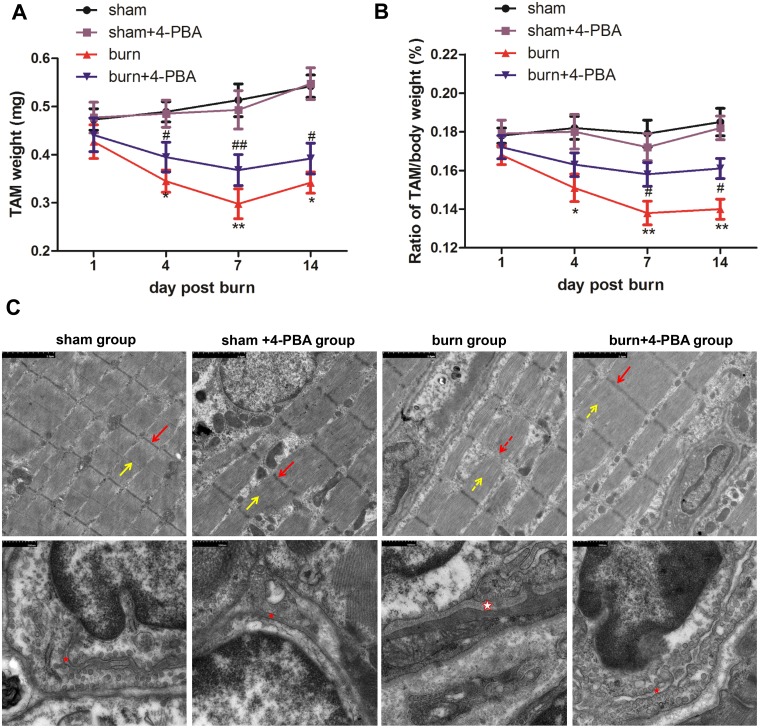
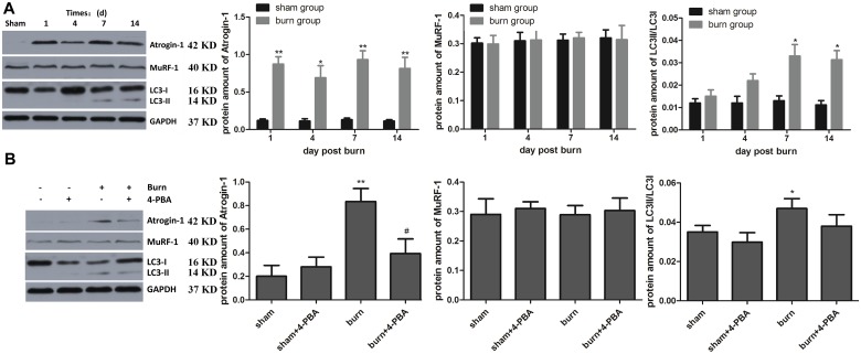
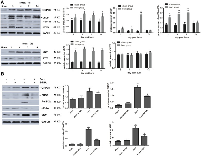
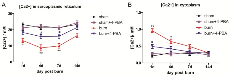
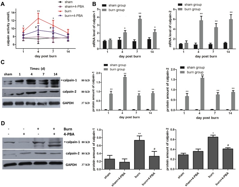
Severe burns are typically followed by hypermetabolism characterized by significant muscle wasting, which causes considerable morbidity and mortality. The aim of the present study was to explore the underlying mechanisms of skeletal muscle damage/wasting post-burn. Rats were randomized to the sham, sham+4-phenylbutyrate (4-PBA, a pharmacological chaperone promoting endoplasmic reticulum (ER) folding/trafficking, commonly considered as an inhibitor of ER), burn (30% total body surface area), and burn+4-PBA groups; and sacrificed at 1, 4, 7, 14 days after the burn injury. Tibial anterior muscle was harvested for transmission electron microscopy, calcium imaging, gene expression and protein analysis of ER stress / ubiquitin-proteasome system / autophagy, and calpain activity measurement. The results showed that ER stress markers were increased in the burn group compared with the sham group, especially at post-burn days 4 and 7, which might consequently elevate cytoplasmic calcium concentration, promote calpain production as well as activation, and cause skeletal muscle damage/wasting of TA muscle after severe burn injury. Interestingly, treatment with 4-PBA prevented burn-induced ER swelling and altered protein expression of ER stress markers and calcium release, attenuating calpain activation and skeletal muscle damage/wasting after severe burn injury. Atrogin-1 and LC3-II/LC3-I ratio were also increased in the burn group compared with the sham group, while MuRF-1 remained unchanged; 4-PBA decreased atrogin-1 in the burn group. Taken together, these findings suggested that severe burn injury induces ER stress, which in turns causes calpain activation. ER stress and subsequent activated calpain play a critical role in skeletal muscle damage/wasting in burned rats.
严重烧伤后通常会出现高代谢,其特征是明显的肌肉萎缩,这会导致相当高的发病率和死亡率。本研究的目的是探讨烧伤后骨骼肌损伤/萎缩的潜在机制。将大鼠随机分为假手术组、假手术+4-苯丁酸盐(4-PBA,一种促进内质网(ER)折叠/运输的药理学伴侣,通常被认为是ER抑制剂)组、烧伤(全身表面积的30%)组和烧伤+4-PBA组;并在烧伤后1、4、7、14天处死。采集胫骨前肌用于透射电子显微镜检查、钙成像、ER应激/泛素-蛋白酶体系统/自噬的基因表达和蛋白质分析以及钙蛋白酶活性测量。结果表明,与假手术组相比,烧伤组的ER应激标志物增加,尤其是在烧伤后第4天和第7天,这可能会因此提高细胞质钙浓度,促进钙蛋白酶的产生以及激活,并导致严重烧伤后TA肌的骨骼肌损伤/萎缩。有趣的是,4-PBA治疗可防止烧伤诱导的ER肿胀,并改变ER应激标志物的蛋白质表达和钙释放,减轻严重烧伤后钙蛋白酶的激活和骨骼肌损伤/萎缩。与假手术组相比,烧伤组的Atrogin-1和LC3-II/LC3-I比值也增加,而MuRF-1保持不变;4-PBA降低了烧伤组的Atrogin-1。综上所述,这些发现表明严重烧伤会诱导ER应激,进而导致钙蛋白酶激活。ER应激和随后激活的钙蛋白酶在烧伤大鼠的骨骼肌损伤/萎缩中起关键作用。