Laboratory of Molecular Biophysics, Department of Cell and Molecular Biology, Uppsala University, Husargatan 3 (Box 596), SE-75124, Uppsala, Sweden.
National Institute for Physiological Sciences (NIPS), Okazaki, Aichi, 444-8585, Japan.
Sci Rep. 2017 Oct 16;7(1):13291. doi: 10.1038/s41598-017-13390-4.
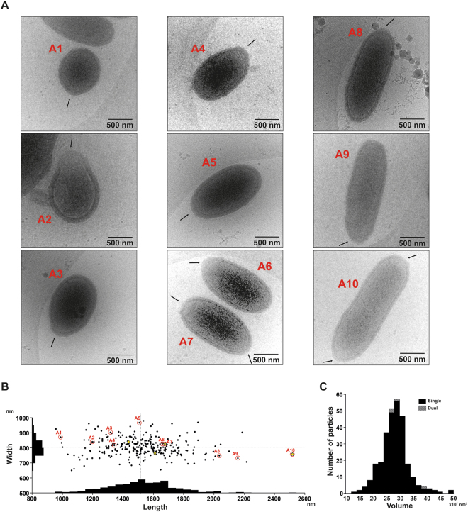
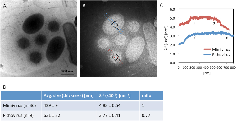
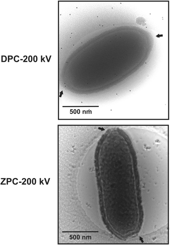
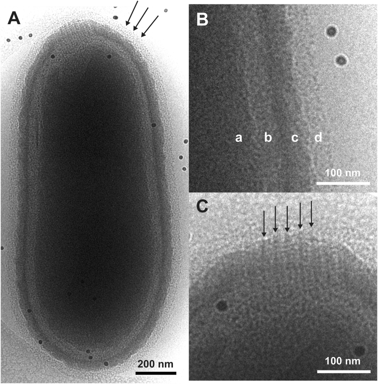
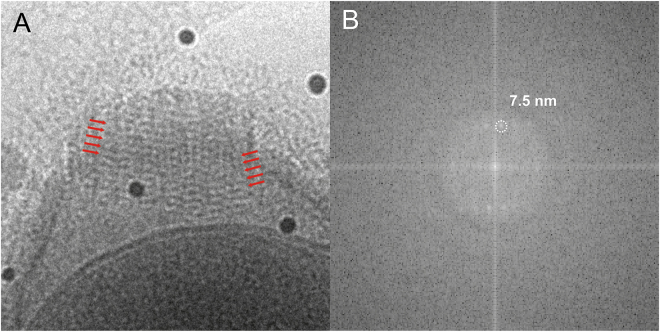
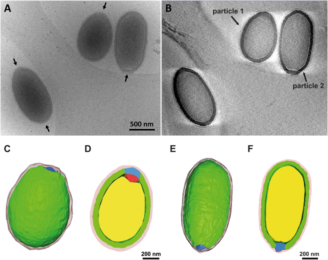
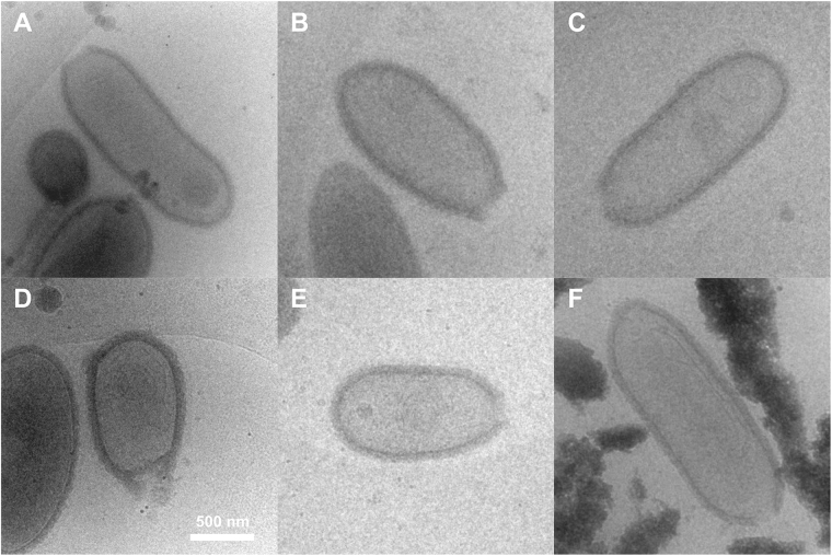
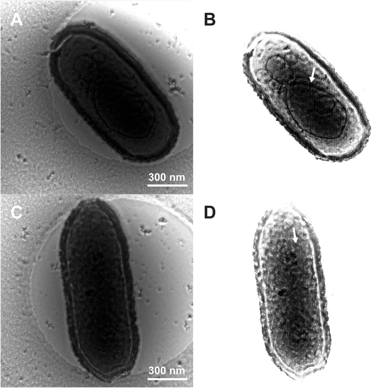
The Pithoviridae giant virus family exhibits the largest viral particle known so far, a prolate spheroid up to 2.5 μm in length and 0.9 μm in diameter. These particles show significant variations in size. Little is known about the structure of the intact virion due to technical limitations with conventional electron cryo-microscopy (cryo-EM) when imaging thick specimens. Here we present the intact structure of the giant Pithovirus sibericum particle at near native conditions using high-voltage electron cryo-tomography (cryo-ET) and energy-filtered cryo-EM. We detected a previously undescribed low-density outer layer covering the tegument and a periodical structuring of the fibres in the striated apical cork. Energy-filtered Zernike phase-contrast cryo-EM images show distinct substructures inside the particles, implicating an internal compartmentalisation. The density of the interior volume of Pithovirus particles is three quarters lower than that of the Mimivirus. However, it is remarkably high given that the 600 kbp Pithovirus genome is only half the size of the Mimivirus genome and is packaged in a volume up to 100 times larger. These observations suggest that the interior is densely packed with macromolecules in addition to the genomic nucleic acid.
潘多拉病毒科的巨型病毒家族展示了目前已知的最大病毒颗粒,其长度可达 2.5μm,直径可达 0.9μm,呈长扁球体。这些颗粒的大小存在显著差异。由于传统电子冷冻显微镜(cryo-EM)在对厚标本成像时存在技术限制,因此对于完整病毒粒子的结构知之甚少。在这里,我们使用高压电子冷冻断层扫描(cryo-ET)和能量过滤冷冻电镜(cryo-EM)在近天然条件下展示了巨型西伯利亚潘多拉病毒粒子的完整结构。我们检测到以前未描述的覆盖外壳的低密度外层,以及条纹状顶端木栓中的纤维的周期性结构。能量过滤的 Zernike 位相对比冷冻电镜图像显示出粒子内部的明显亚结构,暗示内部有隔室化。潘多拉病毒粒子的内部体积密度比 mimivirus 低四分之三。然而,考虑到 600 kbp 的潘多拉病毒基因组仅为 mimivirus 基因组大小的一半,并且包装在大 100 倍的体积中,其密度非常高。这些观察结果表明,除了基因组核酸外,内部还密集地包装了大分子。