Department of Biomedical Engineering, Case Western Reserve University, Cleveland, Ohio, USA.
Department of Pharmacology, Case Western Reserve University, Cleveland, Ohio, USA.
Sci Rep. 2017 Oct 18;7(1):13517. doi: 10.1038/s41598-017-13741-1.
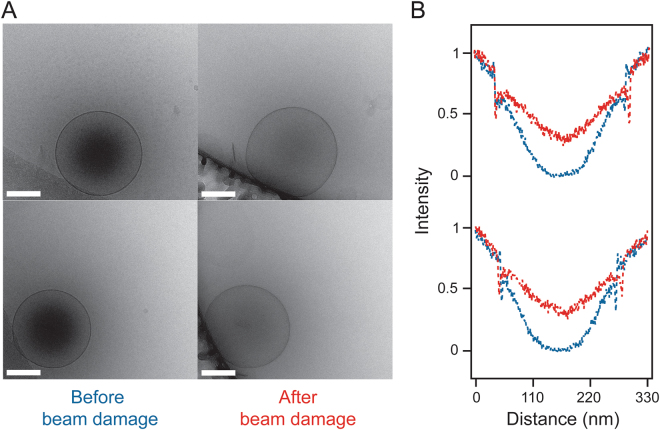
Gas microbubbles stabilized with lipids, surfactants, proteins and/or polymers are widely used clinically as ultrasound contrast agents. Because of their large 1-10 µm size, applications of microbubbles are confined to the blood vessels. Accordingly, there is much interest in generating nanoscale echogenic bubbles (nanobubbles), which can enable new uses of ultrasound contrast agents in molecular imaging and drug delivery, particularly for cancer applications. While the interactions of microbubbles with ultrasound have been widely investigated, little is known about the activity of nanobubbles under ultrasound exposure. In this work, we demonstrate that cryo-electron microscopy (cryo-EM) can be used to image nanoscale lipid and polymer-stabilized perfluorocarbon gas bubbles before and after their destruction with high intensity ultrasound. In addition, cryo-EM can be used to observe electron-beam induced dissipation of nanobubble encapsulated perfluorocarbon gas.
气体微泡通过脂质、表面活性剂、蛋白质和/或聚合物稳定,广泛应用于临床超声造影剂。由于其 1-10μm 的大尺寸,微泡的应用仅限于血管。因此,人们对生成纳米级声敏气泡(纳米气泡)产生了浓厚的兴趣,这可以使超声造影剂在分子成像和药物输送中的新用途成为可能,特别是在癌症应用中。虽然已经广泛研究了微泡与超声的相互作用,但对于纳米气泡在超声照射下的活性知之甚少。在这项工作中,我们证明了冷冻电子显微镜(cryo-EM)可用于在高强度超声破坏前后对纳米级脂质和聚合物稳定的全氟碳气体泡进行成像。此外,cryo-EM 可用于观察电子束诱导纳米气泡包裹的全氟碳气体的耗散。