Dwivedi Durgesh Kumar, Chatzinoff Yonatan, Zhang Yue, Yuan Qing, Fulkerson Michael, Chopra Rajiv, Brugarolas James, Cadeddu Jeffrey A, Kapur Payal, Pedrosa Ivan
Department of Radiology, UT Southwestern Medical Center, Dallas, TX.
Department of Radiology, UT Southwestern Medical Center, Dallas, TX; Department of Advanced Imaging Research Center, UT Southwestern Medical Center, Dallas, TX.
Urology. 2018 Feb;112:209-214. doi: 10.1016/j.urology.2017.08.056. Epub 2017 Oct 19.
To implement a platform for colocalization of in vivo quantitative multiparametric magnetic resonance imaging features with ex vivo surgical specimens of patients with renal masses using patient-specific 3-dimensional (3D)-printed tumor molds, which may aid in targeted tissue procurement and radiomics and radiogenomic analyses.
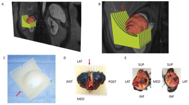
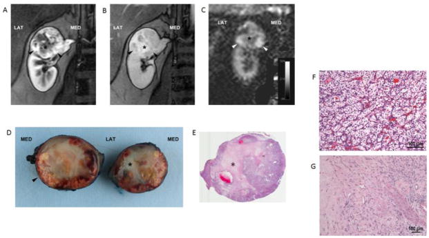
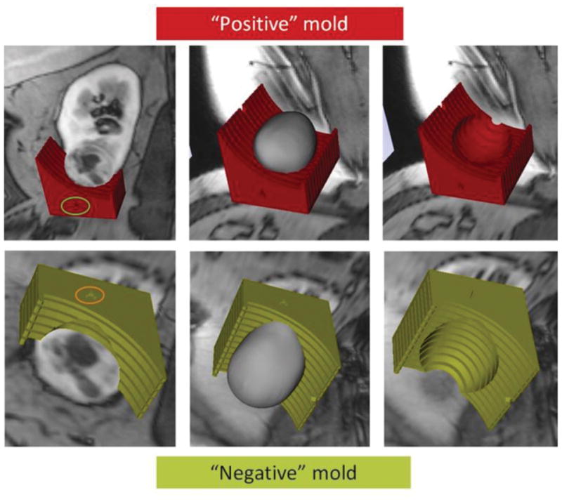
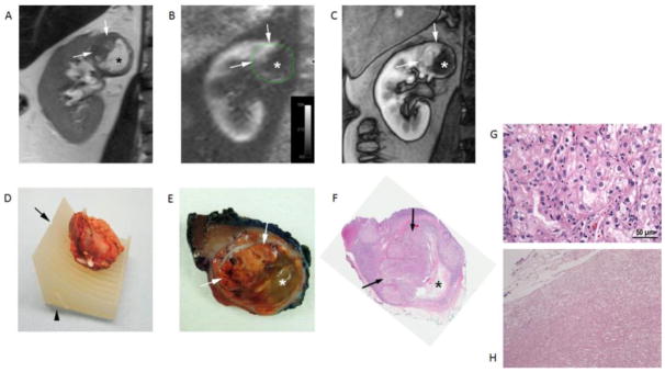
Volumetric segmentation of 6 renal masses was performed with 3D Slicer (http://www.slicer.org) to create a 3D tumor model. A slicing guide template was created with specialized software, which included notches corresponding to the anatomic locations of the magnetic resonance images. The tumor model was subtracted from the slicing guide to create a depression in the slicing guide corresponding to the exact size and shape of the tumor. A customized, tumor-specific, slicing guide was then printed using a 3D printer. After partial nephrectomy, the surgical specimen was bivalved through the preselected magnetic resonance imaging (MRI) plane. A thick slab of the tumor was obtained, fixed, and processed as a whole-mount slide and was correlated to multiparametric MRI findings.
All patients successfully underwent partial nephrectomy and adequate fitting of the tumor specimens within the 3D mold was achieved in all tumors. Distinct in vivo MRI features corresponded to unique pathologic characteristics in the same tumor. The average cost of printing each mold was US$160.7 ± 111.1 (range: US$20.9-$350.7).
MRI-based preoperative 3D printing of tumor-specific molds allow for accurate sectioning of the tumor after surgical resection and colocalization of in vivo imaging features with tissue-based analysis in radiomics and radiogenomic studies.
利用患者特异性三维(3D)打印肿瘤模具,建立一个平台,用于将体内定量多参数磁共振成像特征与肾肿块患者的体外手术标本进行共定位,这可能有助于靶向组织获取以及放射组学和放射基因组学分析。
所有患者均成功接受了部分肾切除术,所有肿瘤的肿瘤标本均能在3D模具中良好适配。同一肿瘤中不同的体内MRI特征对应着独特的病理特征。每个模具的平均打印成本为160.7美元±111.1美元(范围:20.9美元至350.7美元)。
基于MRI的术前肿瘤特异性模具3D打印能够在手术切除后对肿瘤进行精确切片,并在放射组学和放射基因组学研究中实现体内成像特征与基于组织的分析的共定位。