Lee Yeon Hee, Kim Kyoung Nam, Heo Dong Won, Kang Tae Seen, Lee Sung Bok, Kim Chang-Sik
Department of Ophthalmology, Chungnam National University Hospital, Chungnam National University School of Medicine, Daejeon, Korea.
PLoS One. 2017 Oct 26;12(10):e0187093. doi: 10.1371/journal.pone.0187093. eCollection 2017.
To compare the patterns of retinal ganglion cell damage between primary open-angle glaucoma (POAG) and non-arteritic anterior ischaemic optic neuropathy (NAION).
In total, 35 eyes with unilateral NAION, and 70 age- and average peripapillary retinal nerve fibre layer (RNFL) thickness-matched eyes with POAG, were enrolled as disease groups; 35 unaffected fellow eyes of the NAION, and 70 age- and refractive error-matched normal subjects for the POAG, were enrolled as their control groups, respectively. The peripapillary RNFL thickness and macular ganglion cell plus inner plexiform layer (GCIPL) thickness were compared between the disease groups and their controls, and between the two disease groups.
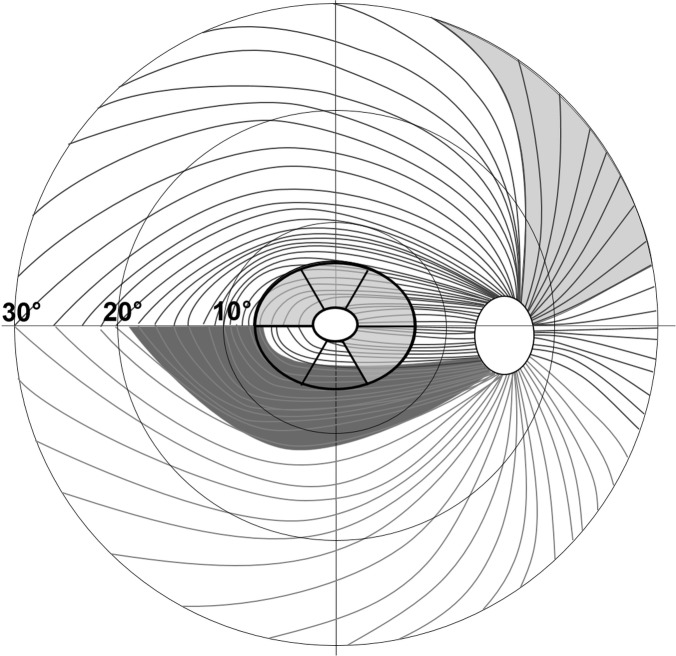
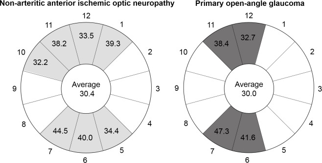
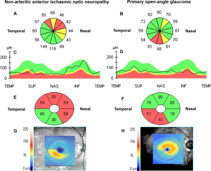
Mean RNFL thicknesses at the 1 and 2 o'clock (superonasal) positions were thinner in NAION than in POAG (both p < 0.05). Mean RNFL thickness at 7 o'clock (inferotemporal) was thinner in POAG than in NAION (p = 0.001). Although there was no significant difference between NAION and POAG in average GCIPL thickness, all of the sectoral GCIPL thicknesses were thinner in NAION (all p < 0.05), except in the inferior and inferotemporal sectors. The ranges of the clock-hour RNFL with damage greater than the average RNFL thickness reduction, versus fellow eyes and control eyes, were 7 hours in NAION and 4 hours in POAG.
The more damaged clock-hour RNFL regions differed between NAION (1 and 2 o'clock) and POAG (7 o'clock). Most sectoral GCIPL thicknesses were thinner in NAION than in POAG.
比较原发性开角型青光眼(POAG)和非动脉炎性前部缺血性视神经病变(NAION)患者视网膜神经节细胞的损伤模式。
将35只单侧NAION患眼和70只年龄及平均视乳头周围视网膜神经纤维层(RNFL)厚度匹配的POAG患眼纳入疾病组;分别将35只NAION患者的健侧眼和70只年龄及屈光不正匹配的POAG正常受试者纳入各自的对照组。比较疾病组与其对照组之间以及两组疾病之间的视乳头周围RNFL厚度和黄斑神经节细胞加内丛状层(GCIPL)厚度。
NAION患者1点和2点(鼻上)位置的平均RNFL厚度比POAG患者薄(均p<0.05)。POAG患者7点(颞下)位置的平均RNFL厚度比NAION患者薄(p = 0.001)。虽然NAION和POAG患者的平均GCIPL厚度无显著差异,但除下方和颞下象限外,NAION患者所有象限的GCIPL厚度均较薄(均p<0.05)。与健侧眼和对照眼相比,RNFL损伤超过平均RNFL厚度减少范围的钟点范围在NAION患者中为7个钟点,在POAG患者中为4个钟点。
NAION(1点和2点)和POAG(7点)患者RNFL损伤更严重的钟点区域不同。NAION患者大多数象限的GCIPL厚度比POAG患者薄。