Department of Pharmacology, Alberta Diabetes Institute, University of Alberta, Edmonton, AB, Canada T6G 2R3.
Center for Biopharmaceuticals, Department of Drug Design and Pharmacology, University of Copenhagen, DK-2100 Copenhagen, Denmark.
Proc Natl Acad Sci U S A. 2017 Nov 7;114(45):E9702-E9711. doi: 10.1073/pnas.1705802114. Epub 2017 Oct 23.
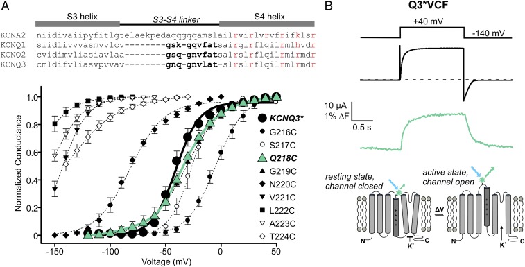
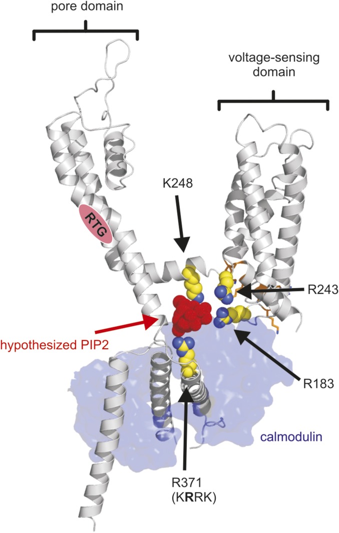
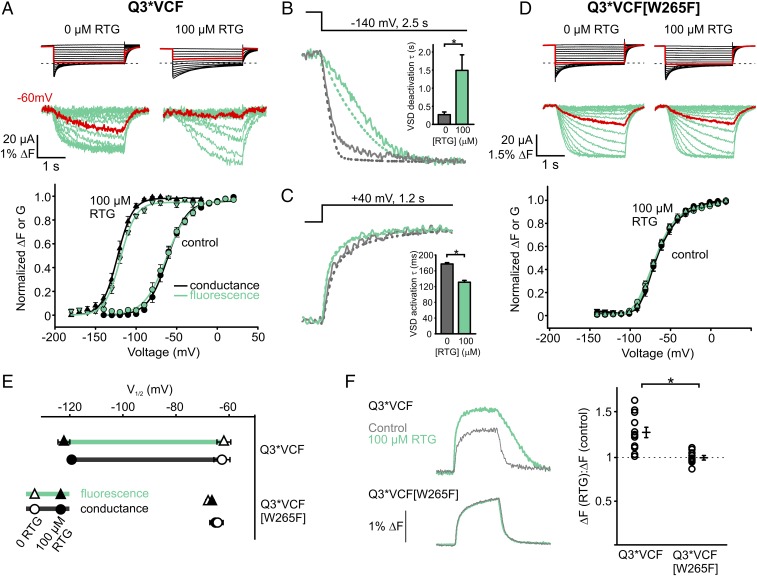
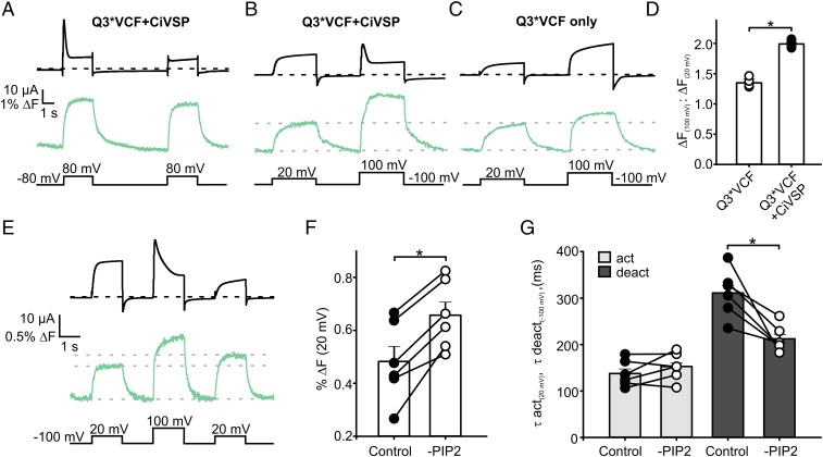
Retigabine (RTG) is a first-in-class antiepileptic drug that suppresses neuronal excitability through the activation of voltage-gated KCNQ2-5 potassium channels. Retigabine binds to the pore-forming domain, causing a hyperpolarizing shift in the voltage dependence of channel activation. To elucidate how the retigabine binding site is coupled to changes in voltage sensing, we used voltage-clamp fluorometry to track conformational changes of the KCNQ3 voltage-sensing domains (VSDs) in response to voltage, retigabine, and PIP2. Steady-state ionic conductance and voltage sensor fluorescence closely overlap under basal PIP2 conditions. Retigabine stabilizes the conducting conformation of the pore and the activated voltage sensor conformation, leading to dramatic deceleration of current and fluorescence deactivation, but these effects are attenuated upon disruption of channel:PIP2 interactions. These findings reveal an important role for PIP2 in coupling retigabine binding to altered VSD function. We identify a polybasic motif in the proximal C terminus of retigabine-sensitive KCNQ channels that contributes to VSD-pore coupling via PIP2, and thereby influences the unique gating effects of retigabine.
瑞替加滨(RTG)是一种首创的抗癫痫药物,通过激活电压门控 KCNQ2-5 钾通道来抑制神经元兴奋性。瑞替加滨与孔形成域结合,导致通道激活的电压依赖性产生超极化偏移。为了阐明瑞替加滨结合位点如何与电压感应变化偶联,我们使用电压钳荧光法跟踪 KCNQ3 电压感应结构域(VSD)在响应电压、瑞替加滨和 PIP2 时的构象变化。在基础 PIP2 条件下,稳态离子电导和电压传感器荧光紧密重叠。瑞替加滨稳定了孔的传导构象和激活的电压传感器构象,导致电流显著减速和荧光失活,但这些效应在破坏通道:PIP2 相互作用时会减弱。这些发现揭示了 PIP2 在将瑞替加滨结合与改变的 VSD 功能偶联中的重要作用。我们确定了瑞替加滨敏感的 KCNQ 通道近端 C 末端的一个多碱性基序,通过 PIP2 有助于 VSD-孔偶联,从而影响瑞替加滨的独特门控效应。