Nalcı Hilal, Şermet Figen, Demirel Sibel, Özmert Emin
Ankara University Faculty of Medicine, Department of Ophthalmology, Ankara, Turkey.
Turk J Ophthalmol. 2017 Oct;47(5):279-284. doi: 10.4274/tjo.68335. Epub 2017 Oct 27.
To evaluate the vascular changes of idiopathic macular telangiectasia type 2 (MacTel 2) patients with optical coherence tomography angiography (OCTA) and correlate these changes with the findings of spectral domain optical coherence tomography (SD-OCT).
Simultaneous SD-OCT and OCTA images of 10 eyes of 6 patients who were diagnosed as MacTel 2 in Ankara University Faculty of Medicine, Department of Ophthalmology were obtained and graded according to the OCTA grading system for MacTel 2.
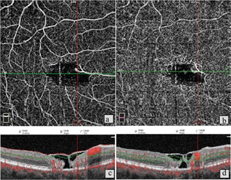
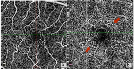
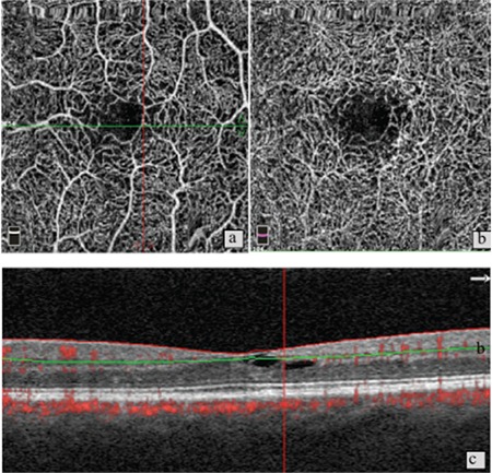
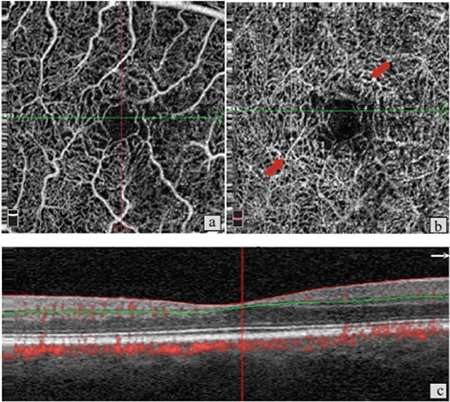
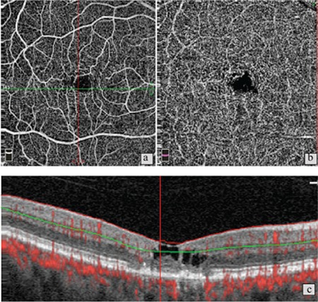
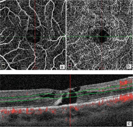
Ten eyes of 6 patients were included. Four (66%) patients were female and 2 (34%) were male. The disease was grade 0 in 2 eyes, grade 1 in 2 eyes, grade 2 in 3 eyes, grade 3 in 1 eye, grade 4 in 1 eye, and grade 5 in 1 eye. The most common findings in grade 1, 2, and 3 non-proliferative disease were thinning of the outer retinal layers, presence of intraretinal hyporeflective layers and inner limiting membrane draping. In cases with subretinal choroidal neovascularisation (CNV) in OCTA, CNV or CNV scar was present in the B-scan SD-OCT images. In a case in which OCT was within normal limits, vascular changes consistent with grade 1 disease were observed in OCTA. On the contrary, 2 patients with significant foveal atrophy and macular hole in B-scan showed changes of early disease in OCTA. In some of the eyes, OCTA revealed an intact superficial vascular layer despite visible changes in the deep layer and the presence of neovascularisation.
OCTA yields findings which are important for understanding the pathogenesis of the disease and providing better follow-up. Contrary to fundus fluorescein angiography, changes in the deep arterial plexus in the early disease and CNV can be clearly observed with OCTA. To achieve the best results in clinical practice, en face flow maps should be evaluated together with B-scan SD-OCT images.
采用光学相干断层扫描血管造影(OCTA)评估2型特发性黄斑毛细血管扩张症(MacTel 2)患者的血管变化,并将这些变化与频域光学相干断层扫描(SD - OCT)的结果相关联。
获取了安卡拉大学医学院眼科诊断为MacTel 2的6例患者10只眼的同步SD - OCT和OCTA图像,并根据MacTel 2的OCTA分级系统进行分级。
纳入6例患者的10只眼。4例(66%)为女性,2例(34%)为男性。2只眼疾病为0级,2只眼为1级,3只眼为2级,1只眼为3级,1只眼为4级,1只眼为5级。1、2和3级非增殖性疾病最常见的表现为视网膜外层变薄、视网膜内低反射层的存在以及内界膜下垂。在OCTA显示有视网膜下脉络膜新生血管(CNV)的病例中,B超SD - OCT图像中存在CNV或CNV瘢痕。在1例OCT正常的病例中,OCTA观察到与1级疾病一致的血管变化。相反,2例B超显示有明显黄斑萎缩和黄斑裂孔的患者在OCTA中表现为早期疾病的变化。在一些眼中,尽管深层有可见变化且存在新生血管,但OCTA显示浅表血管层完整。
OCTA的结果对于理解疾病的发病机制和提供更好的随访很重要。与眼底荧光血管造影不同,OCTA可以清晰观察到早期疾病中深层动脉丛的变化和CNV。为在临床实践中获得最佳结果,应将正面血流图与B超SD - OCT图像一起评估。