• 文献检索
  • 文档翻译
  • 深度研究
  • 学术资讯
  • Suppr Zotero 插件Zotero 插件
  • 邀请有礼
  • 套餐&价格
  • 历史记录
应用&插件
Suppr Zotero 插件Zotero 插件浏览器插件Mac 客户端Windows 客户端微信小程序
定价
高级版会员购买积分包购买API积分包
服务
文献检索文档翻译深度研究API 文档MCP 服务
关于我们
关于 Suppr公司介绍联系我们用户协议隐私条款
关注我们

Suppr 超能文献

核心技术专利:CN118964589B侵权必究
粤ICP备2023148730 号-1Suppr @ 2026

文献检索

告别复杂PubMed语法,用中文像聊天一样搜索,搜遍4000万医学文献。AI智能推荐,让科研检索更轻松。

立即免费搜索

文件翻译

保留排版,准确专业,支持PDF/Word/PPT等文件格式,支持 12+语言互译。

免费翻译文档

深度研究

AI帮你快速写综述,25分钟生成高质量综述,智能提取关键信息,辅助科研写作。

立即免费体验

胶质母细胞瘤成像的标准临床方法和新兴模式

Standard clinical approaches and emerging modalities for glioblastoma imaging.

作者信息

Bernstock Joshua D, Gary Sam E, Klinger Neil, Valdes Pablo A, Ibn Essayed Walid, Olsen Hannah E, Chagoya Gustavo, Elsayed Galal, Yamashita Daisuke, Schuss Patrick, Gessler Florian A, Paolo Peruzzi Pier, Bag Asim K, Friedman Gregory K

机构信息

Department of Neurosurgery, Brigham and Women's Hospital, Harvard Medical School, Boston, Massachusetts, USA.

Medical Scientist Training Program, University of Alabama at Birmingham, Birmingham, Alabama, USA.

出版信息

Neurooncol Adv. 2022 May 26;4(1):vdac080. doi: 10.1093/noajnl/vdac080. eCollection 2022 Jan-Dec.

DOI:10.1093/noajnl/vdac080
PMID:35821676
原文链接:https://pmc.ncbi.nlm.nih.gov/articles/PMC9268747/
Abstract

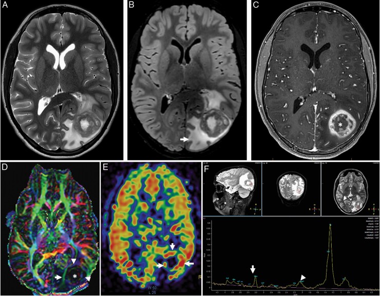
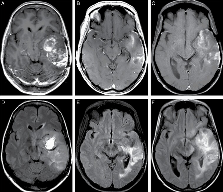
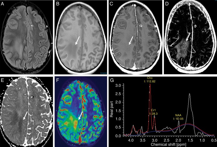
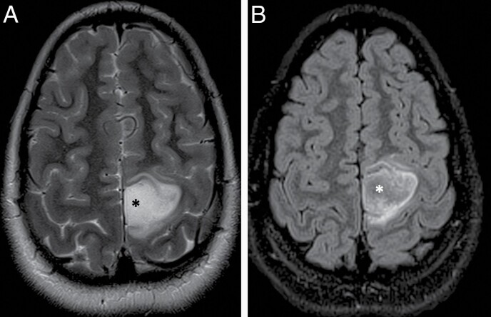
Glioblastoma (GBM) is the most common primary adult intracranial malignancy and carries a dismal prognosis despite an aggressive multimodal treatment regimen that consists of surgical resection, radiation, and adjuvant chemotherapy. Radiographic evaluation, largely informed by magnetic resonance imaging (MRI), is a critical component of initial diagnosis, surgical planning, and post-treatment monitoring. However, conventional MRI does not provide information regarding tumor microvasculature, necrosis, or neoangiogenesis. In addition, traditional MRI imaging can be further confounded by treatment-related effects such as pseudoprogression, radiation necrosis, and/or pseudoresponse(s) that preclude clinicians from making fully informed decisions when structuring a therapeutic approach. A myriad of novel imaging modalities have been developed to address these deficits. Herein, we provide a clinically oriented review of standard techniques for imaging GBM and highlight emerging technologies utilized in disease characterization and therapeutic development.

摘要

胶质母细胞瘤(GBM)是成人最常见的原发性颅内恶性肿瘤,尽管采用了包括手术切除、放疗和辅助化疗在内的积极多模式治疗方案,但其预后仍然很差。主要由磁共振成像(MRI)提供信息的影像学评估是初始诊断、手术规划和治疗后监测的关键组成部分。然而,传统MRI无法提供有关肿瘤微血管系统、坏死或新生血管形成的信息。此外,传统MRI成像可能会因治疗相关效应(如假性进展、放射性坏死和/或假性反应)而进一步混淆,这使得临床医生在制定治疗方案时无法做出充分知情的决策。已经开发了多种新型成像方式来解决这些不足。在此,我们提供了一篇以临床为导向的关于GBM成像标准技术的综述,并强调了用于疾病特征描述和治疗开发的新兴技术。

https://cdn.ncbi.nlm.nih.gov/pmc/blobs/063a/9268747/ecbb5a66b30d/vdac080_fig8.jpg
https://cdn.ncbi.nlm.nih.gov/pmc/blobs/063a/9268747/2350278d86f9/vdac080_fig1.jpg
https://cdn.ncbi.nlm.nih.gov/pmc/blobs/063a/9268747/add85f7b1756/vdac080_fig2.jpg
https://cdn.ncbi.nlm.nih.gov/pmc/blobs/063a/9268747/dd564b770dcc/vdac080_fig3.jpg
https://cdn.ncbi.nlm.nih.gov/pmc/blobs/063a/9268747/346b193a5dde/vdac080_fig4.jpg
https://cdn.ncbi.nlm.nih.gov/pmc/blobs/063a/9268747/8a1cf1e39717/vdac080_fig5.jpg
https://cdn.ncbi.nlm.nih.gov/pmc/blobs/063a/9268747/b38d44b8951d/vdac080_fig6.jpg
https://cdn.ncbi.nlm.nih.gov/pmc/blobs/063a/9268747/8fa0394300cc/vdac080_fig7.jpg
https://cdn.ncbi.nlm.nih.gov/pmc/blobs/063a/9268747/ecbb5a66b30d/vdac080_fig8.jpg

相似文献

1
Standard clinical approaches and emerging modalities for glioblastoma imaging.胶质母细胞瘤成像的标准临床方法和新兴模式
Neurooncol Adv. 2022 May 26;4(1):vdac080. doi: 10.1093/noajnl/vdac080. eCollection 2022 Jan-Dec.
2
Radiation Necrosis, Pseudoprogression, Pseudoresponse, and Tumor Recurrence: Imaging Challenges for the Evaluation of Treated Gliomas.放射性坏死、假性进展、假性缓解和肿瘤复发:治疗后脑胶质瘤评估的影像学挑战。
Contrast Media Mol Imaging. 2018 Dec 2;2018:6828396. doi: 10.1155/2018/6828396. eCollection 2018.
3
Imaging Glioblastoma Posttreatment: Progression, Pseudoprogression, Pseudoresponse, Radiation Necrosis.胶质母细胞瘤治疗后的影像学表现:进展、假性进展、假性反应、放射性坏死
Radiol Clin North Am. 2019 Nov;57(6):1199-1216. doi: 10.1016/j.rcl.2019.07.003. Epub 2019 Aug 16.
4
Combination of IVIM-DWI and 3D-ASL for differentiating true progression from pseudoprogression of Glioblastoma multiforme after concurrent chemoradiotherapy: study protocol of a prospective diagnostic trial.IVIM-DWI与3D-ASL联合用于鉴别多形性胶质母细胞瘤同步放化疗后真性进展与假性进展:一项前瞻性诊断试验的研究方案
BMC Med Imaging. 2017 Feb 1;17(1):10. doi: 10.1186/s12880-017-0183-y.
5
Treatment-related brain tumor imaging changes: So-called "pseudoprogression" vs. tumor progression: Review and future research opportunities.与治疗相关的脑肿瘤影像学变化:所谓的“假性进展”与肿瘤进展:综述及未来研究机遇
Surg Neurol Int. 2013 Apr 17;4(Suppl 3):S129-35. doi: 10.4103/2152-7806.110661. Print 2013.
6
Imaging Glioblastoma Posttreatment: Progression, Pseudoprogression, Pseudoresponse, Radiation Necrosis.脑胶质瘤治疗后影像学表现:进展、假性进展、假性缓解、放射性坏死。
Neuroimaging Clin N Am. 2021 Feb;31(1):103-120. doi: 10.1016/j.nic.2020.09.010.
7
How treatment monitoring is influencing treatment decisions in glioblastomas.治疗监测如何影响胶质母细胞瘤的治疗决策。
Curr Treat Options Neurol. 2015 Apr;17(4):343. doi: 10.1007/s11940-015-0343-8.
8
Advanced magnetic resonance imaging in glioblastoma: a review.胶质母细胞瘤的高级磁共振成像:综述
Chin Clin Oncol. 2017 Aug;6(4):40. doi: 10.21037/cco.2017.06.28.
9
Congress of neurological surgeons systematic review and evidence-based guidelines update on the role of emerging developments in the management of newly diagnosed glioblastoma.神经外科医师大会对新诊断胶质母细胞瘤治疗中新兴进展的作用进行了系统评价和基于证据的指南更新。
J Neurooncol. 2020 Nov;150(2):269-359. doi: 10.1007/s11060-020-03607-4. Epub 2020 Nov 19.
10
Population-based study of pseudoprogression after chemoradiotherapy in GBM.基于人群的胶质母细胞瘤放化疗后假性进展研究。
Can J Neurol Sci. 2009 Sep;36(5):617-22. doi: 10.1017/s0317167100008131.

引用本文的文献

1
Machine Learning-Driven radiomics on 18 F-FDG PET for glioma diagnosis: a systematic review and meta-analysis.基于18F-FDG PET的机器学习驱动的放射组学在胶质瘤诊断中的应用:一项系统评价和荟萃分析
Cancer Imaging. 2025 Aug 26;25(1):106. doi: 10.1186/s40644-025-00915-8.
2
Predictive Digital Twins with Quantified Uncertainty for Patient-Specific Decision Making in Oncology.用于肿瘤学中患者特异性决策的具有量化不确定性的预测性数字孪生模型
ArXiv. 2025 May 13:arXiv:2505.08927v1.
3
Extension of T Hyperintense Areas in Patients With a Glioma: A Comparison Between High-Quality 7 T MRI and Clinical Scans.

本文引用的文献

1
Fundamentals and developments in fluorescence-guided cancer surgery.荧光引导癌症手术的基础与进展
Nat Rev Clin Oncol. 2022 Jan;19(1):9-22. doi: 10.1038/s41571-021-00548-3. Epub 2021 Sep 7.
2
C-Methionine PET for Identification of Pediatric High-Grade Glioma Recurrence.^11^C-蛋氨酸 PET 用于识别小儿高级别胶质瘤复发。
J Nucl Med. 2022 May;63(5):664-671. doi: 10.2967/jnumed.120.261891. Epub 2021 Aug 26.
3
Fluorescence-Guided Surgery in the Surgical Treatment of Gliomas: Past, Present and Future.荧光引导手术在胶质瘤外科治疗中的应用:过去、现在与未来
胶质瘤患者T2高信号区域的扩展:高分辨率7T磁共振成像与临床扫描的比较
NMR Biomed. 2025 Mar;38(3):e5316. doi: 10.1002/nbm.5316.
4
HOTAIR expression as a biomarker in glioblastoma multiforme: a comprehensive meta-analysis of current evidence.长链非编码RNA HOTAIR作为多形性胶质母细胞瘤生物标志物的研究:当前证据的综合荟萃分析
Biomark Med. 2025 Feb;19(4):129-137. doi: 10.1080/17520363.2025.2455925. Epub 2025 Jan 27.
5
A field resource for the glioma cerebrospinal fluid proteome: Impacts of resection and location on biomarker discovery.胶质瘤脑脊液蛋白质组的现场资源:切除和位置对生物标志物发现的影响。
Neuro Oncol. 2025 May 15;27(4):948-962. doi: 10.1093/neuonc/noae277.
6
Clinicopathological Parameters and Immunohistochemical Profiles in Correlation with MRI Characteristics in Glioblastomas.胶质母细胞瘤的临床病理参数及免疫组化特征与MRI表现的相关性
Int J Mol Sci. 2024 Dec 4;25(23):13043. doi: 10.3390/ijms252313043.
7
Enhancing Glioblastoma Resection with NIR Fluorescence Imaging: A Systematic Review.近红外荧光成像增强胶质母细胞瘤切除术:一项系统综述
Cancers (Basel). 2024 Nov 27;16(23):3984. doi: 10.3390/cancers16233984.
8
Radiomics-Based Machine Learning with Natural Gradient Boosting for Continuous Survival Prediction in Glioblastoma.基于影像组学的机器学习与自然梯度提升用于胶质母细胞瘤的连续生存预测
Cancers (Basel). 2024 Oct 26;16(21):3614. doi: 10.3390/cancers16213614.
9
A Comprehensive Review on the Role of MRI in the Assessment of Supratentorial Neoplasms: Comparative Insights Into Adult and Pediatric Cases.关于MRI在幕上肿瘤评估中的作用的综合综述:成人与儿童病例的比较见解
Cureus. 2024 Aug 23;16(8):e67553. doi: 10.7759/cureus.67553. eCollection 2024 Aug.
10
Biosensor-Enhanced Organ-on-a-Chip Models for Investigating Glioblastoma Tumor Microenvironment Dynamics.用于研究胶质母细胞瘤肿瘤微环境动力学的生物传感器增强型器官芯片模型。
Sensors (Basel). 2024 Apr 30;24(9):2865. doi: 10.3390/s24092865.
Cancers (Basel). 2021 Jul 13;13(14):3508. doi: 10.3390/cancers13143508.
4
Fluorescence-Guided Surgery: A Review on Timing and Use in Brain Tumor Surgery.荧光引导手术:脑肿瘤手术中时机与应用的综述
Front Neurol. 2021 Jun 16;12:682151. doi: 10.3389/fneur.2021.682151. eCollection 2021.
5
Challenges and Opportunities of Intraoperative 3D Ultrasound With Neuronavigation in Relation to Intraoperative MRI.术中三维超声与神经导航结合相对于术中磁共振成像的挑战与机遇
Front Oncol. 2021 May 3;11:656519. doi: 10.3389/fonc.2021.656519. eCollection 2021.
6
Oncolytic HSV-1 G207 Immunovirotherapy for Pediatric High-Grade Gliomas.溶瘤单纯疱疹病毒 1 型 G207 免疫病毒疗法治疗儿科高级别脑胶质瘤。
N Engl J Med. 2021 Apr 29;384(17):1613-1622. doi: 10.1056/NEJMoa2024947. Epub 2021 Apr 10.
7
Advanced Imaging Techniques for Radiotherapy Planning of Gliomas.用于胶质瘤放射治疗计划的先进成像技术
Cancers (Basel). 2021 Mar 3;13(5):1063. doi: 10.3390/cancers13051063.
8
Diffusion histology imaging differentiates distinct pediatric brain tumor histology.扩散组织成像可区分不同的小儿脑肿瘤组织学。
Sci Rep. 2021 Feb 26;11(1):4749. doi: 10.1038/s41598-021-84252-3.
9
Applications of indocyanine green in brain tumor surgery: review of clinical evidence and emerging technologies.吲哚菁绿在脑肿瘤手术中的应用:临床证据及新兴技术综述
Neurosurg Focus. 2021 Jan;50(1):E4. doi: 10.3171/2020.10.FOCUS20782.
10
FET PET Radiomics for Differentiating Pseudoprogression from Early Tumor Progression in Glioma Patients Post-Chemoradiation.用于鉴别胶质瘤患者放化疗后假性进展与早期肿瘤进展的FET PET放射组学
Cancers (Basel). 2020 Dec 18;12(12):3835. doi: 10.3390/cancers12123835.