Chaudhury Sidhartha, Ripoll Daniel R, Wallqvist Anders
Department of Defense Biotechnology High Performance Computing Software Applications Institute, Telemedicine and Advanced Technology Research Center, U.S. Army Medical Research and Materiel Command, Ft. Detrick, MD 21702, United States.
Biochem Biophys Rep. 2015 Oct 31;4:375-385. doi: 10.1016/j.bbrep.2015.10.014. eCollection 2015 Dec.
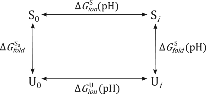
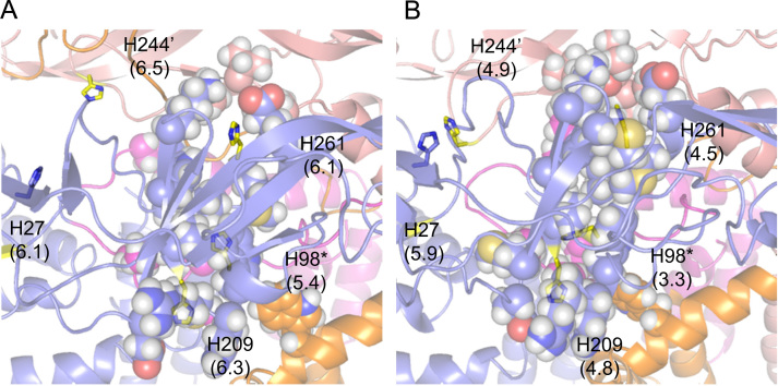
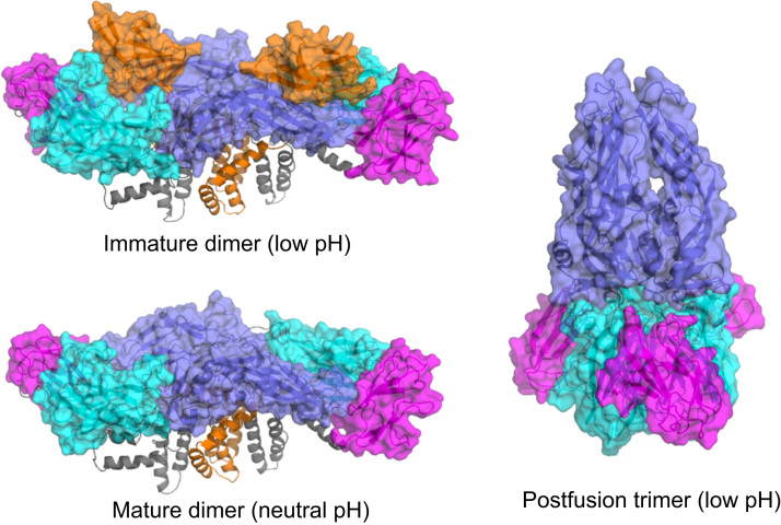
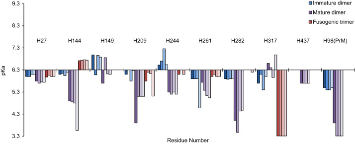
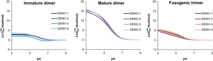
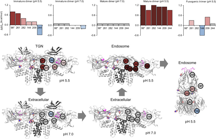
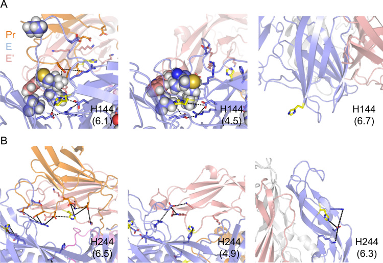
pH-induced conformational changes in dengue virus (DENV) are critical to its ability to infect host cells. The envelope protein heterodimers that make up the viral envelope shift from a dimer to a trimer conformation at low-pH during membrane fusion. Previous studies have suggested that the ionization of histidine residues at low-pH is central to this pH-induced conformational change. We sought out to use molecular modeling with structure-based pKa prediction to provide a quantitative basis for the role of histidines in pH-induced conformational changes and identify which histidine residues were primarily responsible for this transition. We combined existing crystallographic and cryo-electron microscopy data to construct templates of the dimer and trimer conformations for the mature and immature virus. We then generated homology models for the four DENV serotypes and carried out structure-based pKa prediction using Rosetta. Our results showed that the pKa values of a subset of conserved histidines in DENV successfully capture the thermodynamics necessary to drive pH-induced conformational changes during fusion. Here, we identified the structural determinants underlying these pKa values and compare our findings with previous experimental results.
pH诱导的登革病毒(DENV)构象变化对其感染宿主细胞的能力至关重要。在膜融合过程中,构成病毒包膜的包膜蛋白异二聚体在低pH条件下从二聚体构象转变为三聚体构象。先前的研究表明,低pH条件下组氨酸残基的电离是这种pH诱导的构象变化的核心。我们试图利用基于结构的pKa预测进行分子建模,为组氨酸在pH诱导的构象变化中的作用提供定量依据,并确定哪些组氨酸残基主要负责这种转变。我们结合现有的晶体学和冷冻电子显微镜数据,构建成熟和未成熟病毒的二聚体和三聚体构象模板。然后,我们生成了四种登革病毒血清型的同源模型,并使用Rosetta进行基于结构的pKa预测。我们的结果表明,登革病毒中一组保守组氨酸的pKa值成功捕获了在融合过程中驱动pH诱导的构象变化所需的热力学。在此,我们确定了这些pKa值背后的结构决定因素,并将我们的发现与先前的实验结果进行比较。