Department of Medical Ultrasonics, Institute of Diagnostic and Interventional Ultrasound, The First Affiliated Hospital of Sun Yat-Sen University, Guangzhou, China.
Department of Radiology, the First Affiliated Hospital of Sun Yat-Sen University, Guangzhou, China.
Sci Rep. 2017 Nov 13;7(1):15375. doi: 10.1038/s41598-017-15491-6.
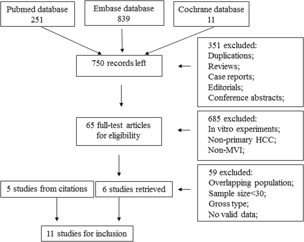
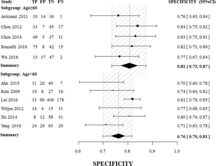
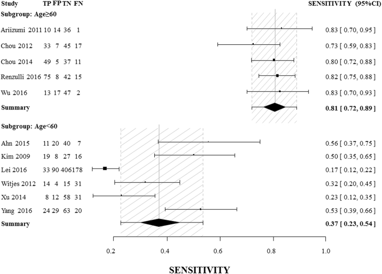
Microvascular invasion (MVI) is rarely diagnosed preoperatively in hepatocellular carcinoma (HCC). The aim of this meta-analysis is to assess the diagnostic power of a non-smooth tumor margin on preoperative imaging for MVI. We performed a literature search using the PubMed, Embase and Cochrane Library databases, and 11 studies were included involving 618 MVI-positive cases and 1030 MVI-negative cases. Considerable heterogeneity was found, and was indicated to be attributable to the mean patient ages in the included studies. In subgroups of studies with a mean patient age older than 60 years and studies with computed tomography (CT) as the imaging method (as opposed to magnetic resonance imaging (MRI)), heterogeneity was low, and the diagnostic odds ratio (DOR) of the single two-dimensional imaging feature for MVI was 21.30 (95% CI [12.52, 36.23]) and 28.78 (95% CI [13.92, 59.36]), respectively; this power was equivalent to or greater than that of certain multivariable-based scoring systems. In conclusion, a non-smooth tumor margin on preoperative imaging is of great value for MVI assessment and should be considered for inclusion in future scoring systems.
微血管侵犯(MVI)在肝细胞癌(HCC)术前很少被诊断。本荟萃分析的目的是评估术前影像学上非平滑肿瘤边缘对 MVI 的诊断能力。我们使用 PubMed、Embase 和 Cochrane 图书馆数据库进行了文献检索,纳入了 11 项研究,共涉及 618 例 MVI 阳性病例和 1030 例 MVI 阴性病例。研究存在较大异质性,表明可能与纳入研究中患者的平均年龄有关。在平均年龄大于 60 岁的亚组研究和以计算机断层扫描(CT)为影像学方法的研究中(而不是磁共振成像(MRI)),异质性较低,MVI 的二维影像学特征的诊断优势比(DOR)分别为 21.30(95%CI[12.52,36.23])和 28.78(95%CI[13.92,59.36]);这种效能相当于或大于某些基于多变量的评分系统。总之,术前影像学上的非平滑肿瘤边缘对 MVI 的评估具有重要价值,应考虑纳入未来的评分系统。