Akahoshi Miho, Abe Koichiro, Uchiyama Yumiko, Momose Mitsuru, Fukushima Kenji, Kitagawa Kazuo, Sakai Shuji
Department of Diagnostic Imaging and Nuclear Medicine Department of Neurology, Tokyo Women's Medical University, Tokyo, Japan.
Medicine (Baltimore). 2017 Nov;96(45):e8484. doi: 10.1097/MD.0000000000008484.
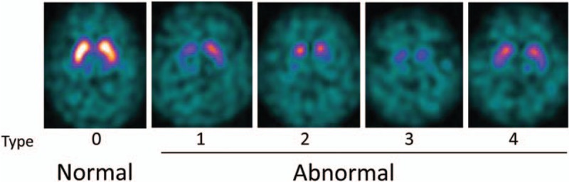
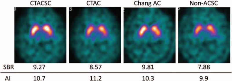
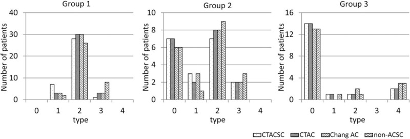
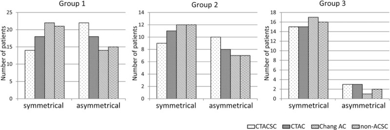
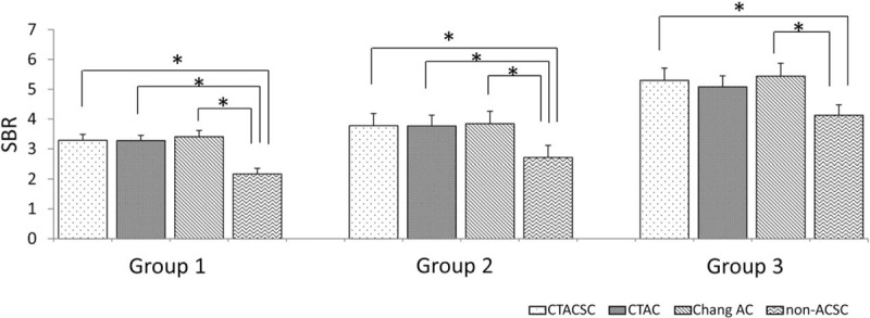
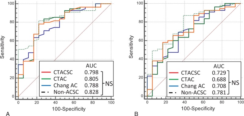
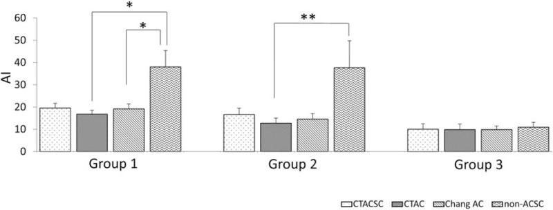
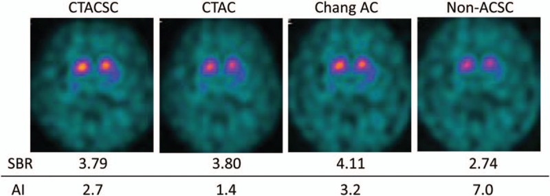
The purpose of this study was to assess the influence of different reconstruction factors in N-ω-fluoropropyl-2β-carbomethoxy-3β-(4-I-123 iodophenyl)nortropane (I-123 FP-CIT) single-photon emission computed tomography (SPECT) images for the diagnosis of dopaminergic system neurodegeneration (DSND).Seventy-three patients (38 females, 35 males) suspected of DSND were included in this study. The patients were divided into 3 groups on the basis of their final clinical diagnoses; patients with Parkinson disease (group 1, n = 36), patients with other DSND (group 2, n = 19), patients without DSND (group 3, n = 18). FP-CIT accumulation in the striata was evaluated visually and semiquantitatively. SPECT images were classified visually as normal or abnormal based on the previous report. For semiquantitative analysis, we used DaTView software (Aze Corporation), and specific binding ratios (SBR) and asymmetry indices (AI) were calculated. Visual and semiquantitative evaluations for different reconstruction factors were compared among the 3 groups.In the visual evaluation, there were no differences among DSND diagnostic capabilities of attenuation and scatter correction by computed tomography attenuation correction scatter correction, computed tomography attenuation correction, Chang attenuation correction, and non-attenuation and -scatter correction. In the semiquantitative evaluation, receiver operating characteristic analysis of SBR and AI for clinical DSND diagnostic ability (group 1+2 vs 3) showed no significant difference among the reconstruction factors by multiple comparisons.Although the values of SBR and AI were changed and image quality could be improved when attenuation correction and/or scatter correction were applied, the clinical impact of these reconstruction factors for the diagnosis of DSND was negligible.
本研究的目的是评估不同重建因素对N-ω-氟丙基-2β-甲氧羰基-3β-(4-I-123碘苯基)去甲托烷(I-123 FP-CIT)单光子发射计算机断层扫描(SPECT)图像诊断多巴胺能系统神经变性(DSND)的影响。本研究纳入了73例疑似DSND的患者(38例女性,35例男性)。根据最终临床诊断将患者分为3组;帕金森病患者(第1组,n = 36),其他DSND患者(第2组,n = 19),无DSND患者(第3组,n = 18)。通过视觉和半定量评估纹状体中FP-CIT的积聚情况。根据先前的报告,将SPECT图像在视觉上分类为正常或异常。对于半定量分析,我们使用DaTView软件(Aze公司),并计算特异性结合率(SBR)和不对称指数(AI)。比较3组之间不同重建因素的视觉和半定量评估结果。在视觉评估中,计算机断层扫描衰减校正散射校正、计算机断层扫描衰减校正、Chang衰减校正以及无衰减和散射校正对DSND的诊断能力之间没有差异。在半定量评估中,通过多重比较,SBR和AI对临床DSND诊断能力(第1组+第2组对比第3组)的受试者工作特征分析显示,各重建因素之间没有显著差异。尽管应用衰减校正和/或散射校正时SBR和AI的值会发生变化且图像质量可以得到改善,但这些重建因素对DSND诊断的临床影响可以忽略不计。