• 文献检索
  • 文档翻译
  • 深度研究
  • 学术资讯
  • Suppr Zotero 插件Zotero 插件
  • 邀请有礼
  • 套餐&价格
  • 历史记录
应用&插件
Suppr Zotero 插件Zotero 插件浏览器插件Mac 客户端Windows 客户端微信小程序
定价
高级版会员购买积分包购买API积分包
服务
文献检索文档翻译深度研究API 文档MCP 服务
关于我们
关于 Suppr公司介绍联系我们用户协议隐私条款
关注我们

Suppr 超能文献

核心技术专利:CN118964589B侵权必究
粤ICP备2023148730 号-1Suppr @ 2026

文献检索

告别复杂PubMed语法,用中文像聊天一样搜索,搜遍4000万医学文献。AI智能推荐,让科研检索更轻松。

立即免费搜索

文件翻译

保留排版,准确专业,支持PDF/Word/PPT等文件格式,支持 12+语言互译。

免费翻译文档

深度研究

AI帮你快速写综述,25分钟生成高质量综述,智能提取关键信息,辅助科研写作。

立即免费体验

弥散加权成像(DWI)与氟代脱氧葡萄糖正电子发射断层显像/X线计算机体层成像(F-FDG PET/CT)在评估胃癌术前N分期中的比较:一项Meta分析的证据

Comparison of DWI and F-FDG PET/CT for assessing preoperative N-staging in gastric cancer: evidence from a meta-analysis.

作者信息

Luo Mingxu, Song Hongmei, Liu Gang, Lin Yikai, Luo Lintao, Zhou Xin, Chen Bo

机构信息

Department of Gastrointestinal Surgery, Xiamen Cancer Hospital, The First Affiliated Hospital of Xiamen University, Xiamen, China.

Department of Oncology, Renmin Hospital of Shiyan, Hubei University of Medicine, Shiyan, China.

出版信息

Oncotarget. 2017 Sep 19;8(48):84473-84488. doi: 10.18632/oncotarget.21055. eCollection 2017 Oct 13.

DOI:10.18632/oncotarget.21055
PMID:29137440
原文链接:https://pmc.ncbi.nlm.nih.gov/articles/PMC5663612/
Abstract

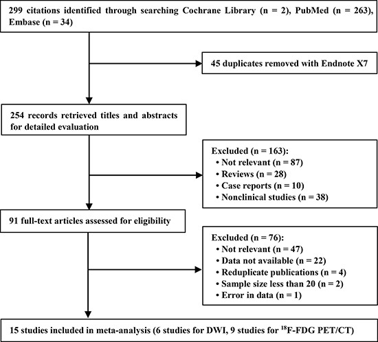
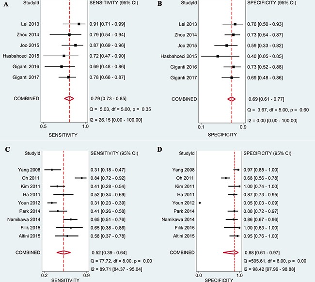
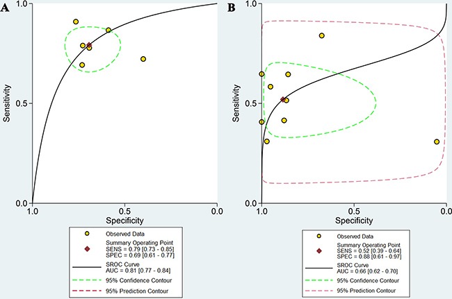
The diagnostic values of diffusion weighted imaging (DWI) and F-fluorodeoxyglucose positron emission tomography/computed tomography (F-FDG PET/CT) for N-staging of gastric cancer (GC) were identified and compared. After a systematic search to identify relevant articles, meta-analysis was used to summarize the sensitivities, specificities, and areas under curves (AUCs) for DWI and PET/CT. To better understand the diagnostic utility of DWI and PET/CT for N-staging, the performance of multi-detector computed tomography (MDCT) was used as a reference. Fifteen studies were analyzed. The pooled sensitivity, specificity, and AUC with 95% confidence intervals of DWI were 0.79 (0.73-0.85), 0.69 (0.61-0.77), and 0.81 (0.77-0.84), respectively. For PET/CT, the corresponding values were 0.52 (0.39-0.64), 0.88 (0.61-0.97), and 0.66 (0.62-0.70), respectively. Comparison of the two techniques revealed DWI had higher sensitivity and AUC, but no difference in specificity. DWI exhibited higher sensitivity but lower specificity than MDCT, and F-FDG PET/CT had lower sensitivity and equivalent specificity. Overall, DWI performed better than F-FDG PET/CT for preoperative N-staging in GC. When the efficacy of MDCT was taken as a reference, DWI represented a complementary imaging technique, while F-FDG PET/CT had limited utility for preoperative N-staging.

摘要

本研究旨在确定并比较弥散加权成像(DWI)和氟脱氧葡萄糖正电子发射断层显像/计算机断层扫描(F-FDG PET/CT)在胃癌(GC)N分期中的诊断价值。在系统检索相关文献后,采用荟萃分析总结DWI和PET/CT的敏感性、特异性及曲线下面积(AUC)。为更好地理解DWI和PET/CT在N分期中的诊断效用,将多排螺旋计算机断层扫描(MDCT)的表现作为参照。共分析了15项研究。DWI的合并敏感性、特异性及95%置信区间的AUC分别为0.79(0.73-0.85)、0.69(0.61-0.77)和0.81(0.77-0.84)。PET/CT的相应值分别为0.52(0.39-0.64)、0.88(0.61-0.97)和0.66(0.62-0.70)。两种技术比较显示,DWI具有更高的敏感性和AUC,但特异性无差异。DWI的敏感性高于MDCT,特异性低于MDCT;F-FDG PET/CT的敏感性较低,特异性相当。总体而言,在GC术前N分期中,DWI的表现优于F-FDG PET/CT。以MDCT的效能为参照时,DWI是一种补充性成像技术,而F-FDG PET/CT在术前N分期中的效用有限。

https://cdn.ncbi.nlm.nih.gov/pmc/blobs/892d/5663612/8c55cf26dbf4/oncotarget-08-84473-g006.jpg
https://cdn.ncbi.nlm.nih.gov/pmc/blobs/892d/5663612/fc713d2542c1/oncotarget-08-84473-g001.jpg
https://cdn.ncbi.nlm.nih.gov/pmc/blobs/892d/5663612/00dffa9abdef/oncotarget-08-84473-g002.jpg
https://cdn.ncbi.nlm.nih.gov/pmc/blobs/892d/5663612/62fd1e5a19f0/oncotarget-08-84473-g003.jpg
https://cdn.ncbi.nlm.nih.gov/pmc/blobs/892d/5663612/d21daa2667ab/oncotarget-08-84473-g004.jpg
https://cdn.ncbi.nlm.nih.gov/pmc/blobs/892d/5663612/09c47ddb68e4/oncotarget-08-84473-g005.jpg
https://cdn.ncbi.nlm.nih.gov/pmc/blobs/892d/5663612/8c55cf26dbf4/oncotarget-08-84473-g006.jpg

相似文献

1
Comparison of DWI and F-FDG PET/CT for assessing preoperative N-staging in gastric cancer: evidence from a meta-analysis.弥散加权成像(DWI)与氟代脱氧葡萄糖正电子发射断层显像/X线计算机体层成像(F-FDG PET/CT)在评估胃癌术前N分期中的比较:一项Meta分析的证据
Oncotarget. 2017 Sep 19;8(48):84473-84488. doi: 10.18632/oncotarget.21055. eCollection 2017 Oct 13.
2
18F-Fluorodeoxyglucose Positron Emission Tomography/Magnetic Resonance in Lymphoma: Comparison With 18F-Fluorodeoxyglucose Positron Emission Tomography/Computed Tomography and With the Addition of Magnetic Resonance Diffusion-Weighted Imaging.18F-氟脱氧葡萄糖正电子发射断层扫描/磁共振成像在淋巴瘤中的应用:与18F-氟脱氧葡萄糖正电子发射断层扫描/计算机断层扫描的比较以及添加磁共振扩散加权成像的研究
Invest Radiol. 2016 Mar;51(3):163-9. doi: 10.1097/RLI.0000000000000218.
3
Preoperative mediastinal and hilar nodal staging with diffusion-weighted magnetic resonance imaging and fluorodeoxyglucose positron emission tomography/computed tomography in patients with non-small-cell lung cancer: which is better?术前纵隔和肺门淋巴结分期的扩散加权磁共振成像与氟代脱氧葡萄糖正电子发射断层扫描/计算机断层扫描在非小细胞肺癌患者中的应用:哪种方法更好?
J Surg Res. 2012 Nov;178(1):304-14. doi: 10.1016/j.jss.2012.03.074. Epub 2012 Apr 22.
4
Preoperative lymph-node staging of invasive urothelial bladder cancer with 18F-fluorodeoxyglucose positron emission tomography/computed axial tomography and magnetic resonance imaging: correlation with histopathology.18F-氟脱氧葡萄糖正电子发射断层扫描/计算机断层扫描和磁共振成像对浸润性膀胱尿路上皮癌的术前淋巴结分期:与组织病理学的相关性
Scand J Urol Nephrol. 2011 Mar;45(2):122-8. doi: 10.3109/00365599.2010.544672. Epub 2011 Jan 13.
5
Diffusion-weighted MR imaging in comparison to integrated [¹⁸F]-FDG PET/CT for N-staging in patients with lung cancer.扩散加权磁共振成像与整合 [¹⁸F]-FDG PET/CT 在肺癌患者 N 分期中的比较。
Eur J Radiol. 2012 Jan;81(1):178-82. doi: 10.1016/j.ejrad.2010.09.001. Epub 2010 Oct 6.
6
Diagnosis of peritoneal dissemination: comparison of 18F-FDG PET/CT, diffusion-weighted MRI, and contrast-enhanced MDCT.腹膜播散的诊断:18F-FDG PET/CT、扩散加权 MRI 和对比增强 MDCT 的比较。
AJR Am J Roentgenol. 2011 Feb;196(2):447-53. doi: 10.2214/AJR.10.4687.
7
F18-fluorodeoxyglucose-positron emission tomography and computed tomography is not accurate in preoperative staging of gastric cancer.F18氟脱氧葡萄糖正电子发射断层扫描与计算机断层扫描在胃癌术前分期中并不准确。
J Korean Surg Soc. 2011 Aug;81(2):104-10. doi: 10.4174/jkss.2011.81.2.104. Epub 2011 Aug 3.
8
Diagnostic value of diffusion-weighted magnetic resonance imaging (DWI) compared to FDG PET/CT for whole-body breast cancer staging.扩散加权磁共振成像(DWI)与 FDG PET/CT 用于全身乳腺癌分期的诊断价值比较。
Eur J Nucl Med Mol Imaging. 2010 Jun;37(6):1077-86. doi: 10.1007/s00259-010-1399-z. Epub 2010 Mar 4.
9
[Evaluation of (18)F-FDG PET/CT in preoperative staging of gastric cancer: a meta-analysis].[18F-FDG PET/CT在胃癌术前分期中的评估:一项荟萃分析]
Zhonghua Wei Chang Wai Ke Za Zhi. 2013 May;16(5):418-24.
10
[Comparison of ¹⁸F-FDG PET/CT and large-scale DWI for evaluation of non-Hodgkin lymphoma bone marrow infiltration].¹⁸F-FDG PET/CT与大规模扩散加权成像用于评估非霍奇金淋巴瘤骨髓浸润的比较
Zhonghua Xue Ye Xue Za Zhi. 2014 Mar;35(3):231-5. doi: 10.3760/cma.j.issn.0253-2727.2014.03.011.

引用本文的文献

1
Chinese national clinical practice guidelines on the prevention, diagnosis, and treatment of early gastric cancer.中国早期胃癌防治国家临床实践指南
Chin Med J (Engl). 2024 Apr 20;137(8):887-908. doi: 10.1097/CM9.0000000000003101. Epub 2024 Mar 21.
2
Diffusion-Weighted Imaging in Oncology: An Update.肿瘤学中的扩散加权成像:最新进展
Cancers (Basel). 2020 Jun 8;12(6):1493. doi: 10.3390/cancers12061493.
3
A Meta-Analysis And Systematic Review Of Accuracy Of Endoscopic Ultrasound For N Staging Of Gastric Cancers.一项关于超声内镜对胃癌N分期准确性的Meta分析与系统评价

本文引用的文献

1
11C-choline PET/CT and whole-body MRI including diffusion-weighted imaging for patients with recurrent prostate cancer.对复发性前列腺癌患者进行11C-胆碱PET/CT及包括弥散加权成像在内的全身MRI检查。
Oncotarget. 2017 Mar 15;8(39):66516-66527. doi: 10.18632/oncotarget.16227. eCollection 2017 Sep 12.
2
Apparent diffusion coefficient by diffusion-weighted magnetic resonance imaging as a sole biomarker for staging and prognosis of gastric cancer.扩散加权磁共振成像的表观扩散系数作为胃癌分期和预后的唯一生物标志物
Chin J Cancer Res. 2017 Apr;29(2):118-126. doi: 10.21147/j.issn.1000-9604.2017.02.04.
3
Contribution of diffusion weighted MRI to diagnosis and staging in gastric tumors and comparison with multi-detector computed tomography.
Cancer Manag Res. 2019 Sep 27;11:8755-8764. doi: 10.2147/CMAR.S200318. eCollection 2019.
4
Gastric cancer and imaging biomarkers: Part 1 - a critical review of DW-MRI and CE-MDCT findings.胃癌与影像学标志物:第一部分——弥散加权 MRI 和 CE-MDCT 研究结果的批判性综述。
Eur Radiol. 2019 Apr;29(4):1743-1753. doi: 10.1007/s00330-018-5732-4. Epub 2018 Oct 2.
5
Is Accurate N - Staging for Gastric Cancer Possible?胃癌能否实现准确的N分期?
Front Surg. 2018 May 31;5:41. doi: 10.3389/fsurg.2018.00041. eCollection 2018.
扩散加权磁共振成像在胃肿瘤诊断及分期中的作用与多排螺旋计算机断层扫描的比较
Radiol Oncol. 2017 Feb 22;51(1):23-29. doi: 10.1515/raon-2017-0002. eCollection 2017 Mar 1.
4
Comparison of 18F-FDG PET/CT and DWI for detection of mediastinal nodal metastasis in non-small cell lung cancer: A meta-analysis.18F-FDG PET/CT与扩散加权成像在检测非小细胞肺癌纵隔淋巴结转移中的比较:一项荟萃分析
PLoS One. 2017 Mar 2;12(3):e0173104. doi: 10.1371/journal.pone.0173104. eCollection 2017.
5
Comparison of Contrast-Enhanced CT and [F]FDG PET/CT Analysis Using Kurtosis and Skewness in Patients with Primary Colorectal Cancer.原发性结直肠癌患者的对比增强 CT 和 [F]FDG PET/CT 分析中使用峰度和偏度的比较。
Mol Imaging Biol. 2017 Oct;19(5):795-803. doi: 10.1007/s11307-017-1066-x.
6
Cancer Statistics, 2017.《2017 年癌症统计》
CA Cancer J Clin. 2017 Jan;67(1):7-30. doi: 10.3322/caac.21387. Epub 2017 Jan 5.
7
PI-RADS version 2: quantitative analysis aids reliable interpretation of diffusion-weighted imaging for prostate cancer.前列腺影像报告和数据系统(PI-RADS)第2版:定量分析有助于对前列腺癌的扩散加权成像进行可靠解读。
Eur Radiol. 2017 Jul;27(7):2776-2783. doi: 10.1007/s00330-016-4678-7. Epub 2016 Dec 12.
8
Predictive Role of the Number of 18F-FDG-Positive Lymph Nodes Detected by PET/CT for Pre-Treatment Evaluation of Locally Advanced Gastric Cancer.PET/CT检测的18F-FDG阳性淋巴结数量对局部进展期胃癌治疗前评估的预测作用
PLoS One. 2016 Dec 9;11(12):e0166836. doi: 10.1371/journal.pone.0166836. eCollection 2016.
9
Can Diffusion-weighted Magnetic Resonance Imaging Predict Survival in Patients with Cervical Cancer? A Meta-Analysis.扩散加权磁共振成像能否预测宫颈癌患者的生存情况?一项荟萃分析。
Eur J Radiol. 2016 Dec;85(12):2174-2181. doi: 10.1016/j.ejrad.2016.10.011. Epub 2016 Oct 13.
10
Application of texture analysis based on apparent diffusion coefficient maps in discriminating different stages of rectal cancer.基于表观扩散系数图的纹理分析在鉴别直肠癌不同分期中的应用。
J Magn Reson Imaging. 2017 Jun;45(6):1798-1808. doi: 10.1002/jmri.25460. Epub 2016 Sep 22.