• 文献检索
  • 文档翻译
  • 深度研究
  • 学术资讯
  • Suppr Zotero 插件Zotero 插件
  • 邀请有礼
  • 套餐&价格
  • 历史记录
应用&插件
Suppr Zotero 插件Zotero 插件浏览器插件Mac 客户端Windows 客户端微信小程序
定价
高级版会员购买积分包购买API积分包
服务
文献检索文档翻译深度研究API 文档MCP 服务
关于我们
关于 Suppr公司介绍联系我们用户协议隐私条款
关注我们

Suppr 超能文献

核心技术专利:CN118964589B侵权必究
粤ICP备2023148730 号-1Suppr @ 2026

文献检索

告别复杂PubMed语法,用中文像聊天一样搜索,搜遍4000万医学文献。AI智能推荐,让科研检索更轻松。

立即免费搜索

文件翻译

保留排版,准确专业,支持PDF/Word/PPT等文件格式,支持 12+语言互译。

免费翻译文档

深度研究

AI帮你快速写综述,25分钟生成高质量综述,智能提取关键信息,辅助科研写作。

立即免费体验

胃癌与影像学标志物:第一部分——弥散加权 MRI 和 CE-MDCT 研究结果的批判性综述。

Gastric cancer and imaging biomarkers: Part 1 - a critical review of DW-MRI and CE-MDCT findings.

机构信息

Department of Radiology, University College London Hospital NHS Foundation Trust, London, UK.

Division of Surgery and Interventional Science, Faculty of Medical Sciences, University College London, 3rd Floor, Charles Bell House, 43-45 Foley St, London, W1W 7TS, UK.

出版信息

Eur Radiol. 2019 Apr;29(4):1743-1753. doi: 10.1007/s00330-018-5732-4. Epub 2018 Oct 2.

DOI:10.1007/s00330-018-5732-4
PMID:30280246
原文链接:https://pmc.ncbi.nlm.nih.gov/articles/PMC6420485/
Abstract

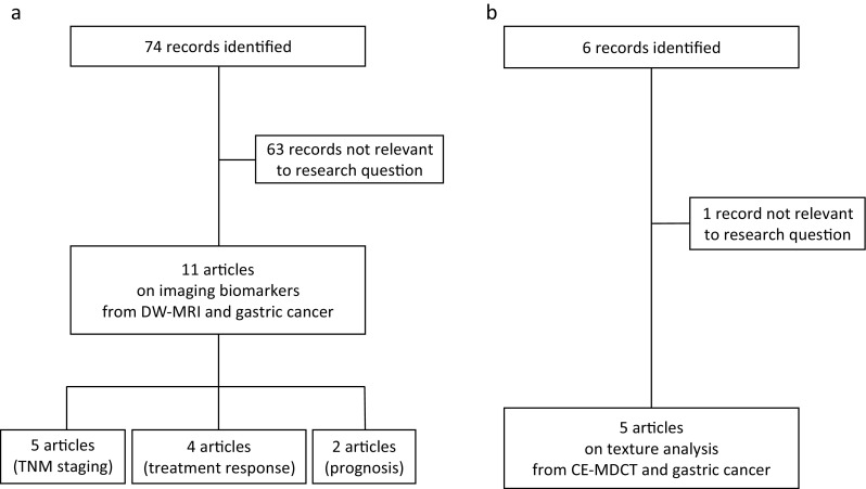
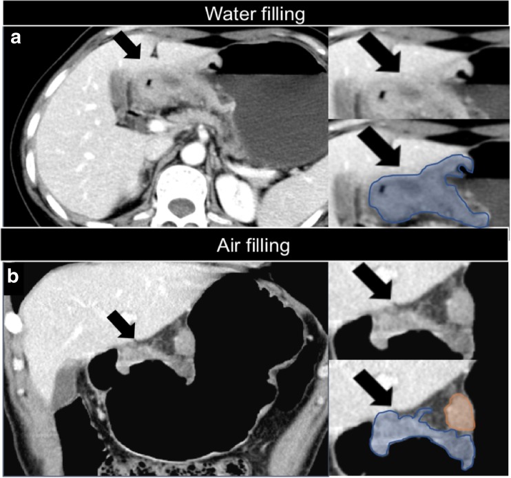
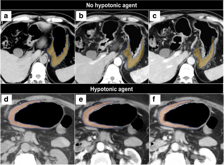
The current standard of care for gastric cancer imaging includes heterogeneity in image acquisition techniques and qualitative image interpretation. In addition to qualitative assessment, several imaging techniques, including diffusion-weighted magnetic resonance imaging (DW-MRI), contrast-enhanced multidetector computed tomography (CE-MDCT), dynamic-contrast enhanced MRI and 18F-fluorodeoxyglucose positron emission tomography, can allow quantitative analysis. However, so far there is no consensus regarding the application of functional imaging in the management of gastric cancer. The aim of this article is to specifically review two promising biomarkers for gastric cancer with reasonable spatial resolution: the apparent diffusion coefficient (ADC) from DW-MRI and textural features from CE-MDCT. We searched MEDLINE/ PubMed for manuscripts published from inception to 6 February 2018. Initially, we searched for (gastric cancer OR gastric tumour) AND diffusion weighted magnetic resonance imaging. Then, we searched for (gastric cancer OR gastric tumour) AND texture analysis AND computed tomography. We collated the results from the studies related to this query. There is evidence that: (1) the ADC is a promising biomarker for the evaluation of the aggressiveness (T and N stage), treatment response and prognosis of gastric cancer; (2) textural features are related to the degree of differentiation, Lauren classification, treatment response and prognosis of gastric cancer. We conclude that these imaging biomarkers hold promise as effective additional tools in the diagnostic pathway of gastric cancer and may facilitate the multidisciplinary work between the radiologist and clinician, and across different institutions, to provide a greater biological understanding of gastric cancer. KEY POINTS: • Quantitative imaging is the extraction of quantifiable features from medical images for the assessment of normal or pathological conditions and represents a promising area for gastric cancer. • Quantitative analysis from CE-MDCT and DW-MRI allows the extrapolation of multiple imaging biomarkers. • ADC from DW-MRI and CE- MDCT-based texture features are non-invasive, quantitative imaging biomarkers that hold promise in the evaluation of the aggressiveness, treatment response and prognosis of gastric cancer.

摘要

当前胃癌影像学的标准治疗包括图像采集技术和定性图像解释的异质性。除了定性评估外,几种影像学技术,包括扩散加权磁共振成像(DW-MRI)、多排螺旋 CT 增强扫描(CE-MDCT)、动态对比增强 MRI 和 18F-氟代脱氧葡萄糖正电子发射断层扫描,都可以进行定量分析。然而,到目前为止,在胃癌的管理中,功能性成像的应用还没有达成共识。本文的目的是专门回顾两种具有合理空间分辨率的有前途的胃癌生物标志物:来自 DW-MRI 的表观扩散系数(ADC)和来自 CE-MDCT 的纹理特征。我们在 MEDLINE/PubMed 上搜索了从开始到 2018 年 2 月 6 日发表的手稿。最初,我们搜索了(胃癌或胃肿瘤)和扩散加权磁共振成像。然后,我们搜索了(胃癌或胃肿瘤)和纹理分析和计算机断层扫描。我们整理了与该查询相关的研究结果。有证据表明:(1)ADC 是评估胃癌侵袭性(T 和 N 期)、治疗反应和预后的有前途的生物标志物;(2)纹理特征与胃癌的分化程度、Lauren 分类、治疗反应和预后有关。我们得出的结论是,这些影像学生物标志物有望成为胃癌诊断途径中的有效辅助工具,并能促进放射科医生和临床医生之间以及不同机构之间的多学科合作,为胃癌提供更深入的生物学认识。要点: • 定量成像技术是从医学图像中提取可量化特征,用于评估正常或病理状况,是胃癌研究的一个有前途的领域。 • CE-MDCT 和 DW-MRI 的定量分析允许推断出多种成像生物标志物。 • DW-MRI 的 ADC 和基于 CE-MDCT 的纹理特征是无创的、定量的成像生物标志物,在评估胃癌的侵袭性、治疗反应和预后方面具有潜力。

https://cdn.ncbi.nlm.nih.gov/pmc/blobs/0d8f/6420485/f955f1d50c3c/330_2018_5732_Fig6_HTML.jpg
https://cdn.ncbi.nlm.nih.gov/pmc/blobs/0d8f/6420485/72747fc059f2/330_2018_5732_Fig1_HTML.jpg
https://cdn.ncbi.nlm.nih.gov/pmc/blobs/0d8f/6420485/24b58ba58d01/330_2018_5732_Fig2_HTML.jpg
https://cdn.ncbi.nlm.nih.gov/pmc/blobs/0d8f/6420485/9a4a65e111c7/330_2018_5732_Fig3_HTML.jpg
https://cdn.ncbi.nlm.nih.gov/pmc/blobs/0d8f/6420485/8b6f011fc772/330_2018_5732_Fig4_HTML.jpg
https://cdn.ncbi.nlm.nih.gov/pmc/blobs/0d8f/6420485/872cd5ca596a/330_2018_5732_Fig5_HTML.jpg
https://cdn.ncbi.nlm.nih.gov/pmc/blobs/0d8f/6420485/f955f1d50c3c/330_2018_5732_Fig6_HTML.jpg

相似文献

1
Gastric cancer and imaging biomarkers: Part 1 - a critical review of DW-MRI and CE-MDCT findings.胃癌与影像学标志物:第一部分——弥散加权 MRI 和 CE-MDCT 研究结果的批判性综述。
Eur Radiol. 2019 Apr;29(4):1743-1753. doi: 10.1007/s00330-018-5732-4. Epub 2018 Oct 2.
2
Gastric cancer and image-derived quantitative parameters: Part 2-a critical review of DCE-MRI and F-FDG PET/CT findings.胃癌与影像衍生定量参数:第二部分——DCE-MRI 和 F-FDG PET/CT 研究结果的批判性综述。
Eur Radiol. 2020 Jan;30(1):247-260. doi: 10.1007/s00330-019-06370-x. Epub 2019 Aug 7.
3
Response to chemotherapy in gastric adenocarcinoma with diffusion-weighted MRI and (18) F-FDG-PET/CT: correlation of apparent diffusion coefficient and partial volume corrected standardized uptake value with histological tumor regression grade.弥散加权磁共振成像和(18)F-FDG-PET/CT评估胃腺癌化疗疗效:表观扩散系数和部分容积校正标准化摄取值与组织学肿瘤退缩分级的相关性
J Magn Reson Imaging. 2014 Nov;40(5):1147-57. doi: 10.1002/jmri.24464. Epub 2013 Nov 8.
4
Use of Diffusion-Weighted Magnetic Resonance Imaging and Apparent Diffusion Coefficient in Gastric Cancer Staging.扩散加权磁共振成像及表观扩散系数在胃癌分期中的应用。
Curr Med Imaging. 2020;16(10):1278-1289. doi: 10.2174/1573405616666200218124926.
5
Upper abdominal gadoxetic acid-enhanced and diffusion-weighted MRI for the detection of gastric cancer: Comparison with two-dimensional multidetector row CT.上腹部钆塞酸增强及扩散加权磁共振成像用于胃癌检测:与二维多排探测器CT的比较
Clin Radiol. 2014 Aug;69(8):827-35. doi: 10.1016/j.crad.2014.03.017. Epub 2014 May 16.
6
Evaluation of the Added Value of Diffusion-Weighted Imaging to Conventional Magnetic Resonance Imaging in Pancreatic Neuroendocrine Tumors and Comparison With 68Ga-DOTANOC Positron Emission Tomography/Computed Tomography.扩散加权成像对胰腺神经内分泌肿瘤常规磁共振成像的附加值评估及与68Ga-DOTANOC正电子发射断层扫描/计算机断层扫描的比较
Pancreas. 2016 Mar;45(3):345-54. doi: 10.1097/MPA.0000000000000461.
7
Preoperative T and N staging of gastric cancer: magnetic resonance imaging (MRI) versus multi detector computed tomography (MDCT).胃癌的术前T和N分期:磁共振成像(MRI)与多层螺旋计算机断层扫描(MDCT)的比较
Clin Ter. 2010;161(2):e57-62.
8
The added value of PET/Ce-CT/DW-MRI fusion in assessment of hepatic focal lesions: PET/Ce-CT/DW-MRI fusion in hepatic focal lesion.PET/Ce-CT/DW-MRI融合成像在肝脏局灶性病变评估中的附加价值:肝脏局灶性病变的PET/Ce-CT/DW-MRI融合成像
Nucl Med Biol. 2015 Jul;42(7):637-42. doi: 10.1016/j.nucmedbio.2015.03.010. Epub 2015 Apr 6.
9
Diffusion-weighted magnetic resonance imaging in the depiction of gastric cancer: initial experience.磁共振弥散加权成像在胃癌中的应用:初步经验。
Abdom Radiol (NY). 2016 Jan;41(1):2-9. doi: 10.1007/s00261-015-0594-6.
10
Imaging the early response to chemotherapy in advanced lung cancer with diffusion-weighted magnetic resonance imaging compared to fluorine-18 fluorodeoxyglucose positron emission tomography and computed tomography.采用弥散加权磁共振成像与氟-18 氟代脱氧葡萄糖正电子发射断层扫描和计算机断层扫描对比评估晚期肺癌化疗的早期疗效。
J Magn Reson Imaging. 2013 Jul;38(1):80-8. doi: 10.1002/jmri.23959. Epub 2012 Dec 12.

引用本文的文献

1
CT-derived extracellular volume fraction as a predictive marker for postoperative recurrence in pStage II-III gastric cancer.CT衍生的细胞外体积分数作为II-III期胃癌术后复发的预测标志物。
Eur Radiol. 2025 Jun 18. doi: 10.1007/s00330-025-11765-0.
2
Multi-orientation and prone-position ZOOMit diffusion-weighted imaging for predicting T stage in distal gastric cancer.多方位及俯卧位ZOOMit扩散加权成像用于预测远端胃癌的T分期
Eur Radiol. 2025 Apr 30. doi: 10.1007/s00330-025-11638-6.
3
Prognostic value of diffusion-weighted imaging to cytoreductive surgery with or without hyperthermic intraperitoneal chemotherapy for patients with gastric cancer and peritoneal metastases.

本文引用的文献

1
Feasibility of free-breathing dynamic contrast-enhanced MRI of gastric cancer using a golden-angle radial stack-of-stars VIBE sequence: comparison with the conventional contrast-enhanced breath-hold 3D VIBE sequence.基于黄金角放射状星型扰相容积内插序列动态对比增强 MRI 对胃癌可行性的研究:与常规对比增强屏气 3D 容积内插扰相序列的比较。
Eur Radiol. 2018 May;28(5):1891-1899. doi: 10.1007/s00330-017-5193-1. Epub 2017 Dec 19.
2
Comparison of DWI and F-FDG PET/CT for assessing preoperative N-staging in gastric cancer: evidence from a meta-analysis.弥散加权成像(DWI)与氟代脱氧葡萄糖正电子发射断层显像/X线计算机体层成像(F-FDG PET/CT)在评估胃癌术前N分期中的比较:一项Meta分析的证据
Oncotarget. 2017 Sep 19;8(48):84473-84488. doi: 10.18632/oncotarget.21055. eCollection 2017 Oct 13.
3
弥散加权成像对接受或未接受热灌注腹腔化疗的胃癌伴腹膜转移患者减瘤手术的预后价值
BMC Cancer. 2025 Apr 5;25(1):616. doi: 10.1186/s12885-025-14008-z.
4
Diffusion-weighted MRI of advanced gastric cancer: correlations of the apparent diffusion coefficient with Borrmann classification, proliferation and aggressiveness.进展期胃癌的扩散加权磁共振成像:表观扩散系数与Borrmann分型、增殖及侵袭性的相关性
Abdom Radiol (NY). 2025 Jan 16. doi: 10.1007/s00261-024-04718-6.
5
Hepatoid adenocarcinoma of the stomach: discrimination from conventional gastric adenocarcinoma with a computed tomography-based radiomics nomogram.胃肝样腺癌:基于计算机断层扫描的影像组学列线图鉴别诊断与传统胃腺癌
J Gastrointest Oncol. 2024 Oct 31;15(5):2041-2052. doi: 10.21037/jgo-24-210. Epub 2024 Oct 25.
6
Predicting tumor invasion depth in gastric cancer: developing and validating multivariate models incorporating preoperative IVIM-DWI parameters and MRI morphological characteristics.预测胃癌肿瘤侵袭深度:建立和验证包含术前 IVIM-DWI 参数和 MRI 形态学特征的多变量模型。
Eur J Med Res. 2024 Aug 22;29(1):431. doi: 10.1186/s40001-024-02017-w.
7
Advanced Gastric Cancer: CT Radiomics Prediction of Lymph Modes Metastasis After Neoadjuvant Chemotherapy.进展期胃癌:新辅助化疗后淋巴结转移的CT影像组学预测
J Imaging Inform Med. 2024 Dec;37(6):2910-2919. doi: 10.1007/s10278-024-01148-0. Epub 2024 Jun 17.
8
A nomogram model of spectral CT quantitative parameters and clinical characteristics predicting lymphovascular invasion of gastric cancer.预测胃癌淋巴管侵犯的光谱CT定量参数与临床特征列线图模型
Heliyon. 2024 Apr 3;10(7):e29214. doi: 10.1016/j.heliyon.2024.e29214. eCollection 2024 Apr 15.
9
Predictive nomogram for lymph node metastasis and survival in gastric cancer using contrast-enhanced computed tomography-based radiomics: a retrospective study.基于增强 CT 影像组学的胃癌淋巴结转移和生存预测列线图:一项回顾性研究。
PeerJ. 2024 Mar 20;12:e17111. doi: 10.7717/peerj.17111. eCollection 2024.
10
Spectral CT-based nomogram for preoperative prediction of perineural invasion in locally advanced gastric cancer: a prospective study.基于光谱 CT 的局部进展期胃癌术前预测神经周围侵犯的列线图:一项前瞻性研究。
Eur Radiol. 2023 Jul;33(7):5172-5183. doi: 10.1007/s00330-023-09464-9. Epub 2023 Feb 24.
Principles of Quantitative MR Imaging with Illustrated Review of Applicable Modular Pulse Diagrams.定量磁共振成像原理及适用模块脉冲图实例复习。
Radiographics. 2017 Nov-Dec;37(7):2083-2105. doi: 10.1148/rg.2017160099. Epub 2017 Oct 6.
4
Whole-lesion apparent diffusion coefficient histogram analysis: significance in T and N staging of gastric cancers.全病灶表观弥散系数直方图分析:在胃癌 T 和 N 分期中的意义。
BMC Cancer. 2017 Oct 2;17(1):665. doi: 10.1186/s12885-017-3622-9.
5
Implementing diffusion-weighted MRI for body imaging in prospective multicentre trials: current considerations and future perspectives.前瞻性多中心试验中体部成像的扩散加权 MRI 的实施:当前考虑因素和未来展望。
Eur Radiol. 2018 Mar;28(3):1118-1131. doi: 10.1007/s00330-017-4972-z. Epub 2017 Sep 27.
6
CT Texture Analysis: Definitions, Applications, Biologic Correlates, and Challenges.CT纹理分析:定义、应用、生物学关联及挑战
Radiographics. 2017 Sep-Oct;37(5):1483-1503. doi: 10.1148/rg.2017170056.
7
Application of CT texture analysis in predicting histopathological characteristics of gastric cancers.CT 纹理分析在预测胃癌组织病理学特征中的应用。
Eur Radiol. 2017 Dec;27(12):4951-4959. doi: 10.1007/s00330-017-4881-1. Epub 2017 Jun 22.
8
Imaging biomarkers exist and they underpin clinical decision-making.影像生物标志物确实存在,并且它们是临床决策的基础。
Nat Rev Clin Oncol. 2017 Jul;14(7):452. doi: 10.1038/nrclinonc.2017.84. Epub 2017 Jun 13.
9
Pre-treatment MDCT-based texture analysis for therapy response prediction in gastric cancer: Comparison with tumour regression grade at final histology.基于治疗前多层螺旋CT的纹理分析在胃癌治疗反应预测中的应用:与最终组织学肿瘤退缩分级的比较
Eur J Radiol. 2017 May;90:129-137. doi: 10.1016/j.ejrad.2017.02.043. Epub 2017 Mar 1.
10
Apparent diffusion coefficient by diffusion-weighted magnetic resonance imaging as a sole biomarker for staging and prognosis of gastric cancer.扩散加权磁共振成像的表观扩散系数作为胃癌分期和预后的唯一生物标志物
Chin J Cancer Res. 2017 Apr;29(2):118-126. doi: 10.21147/j.issn.1000-9604.2017.02.04.