Yang Yiming, Wu Ke, Liu Yulin, Shi Liang, Tao Kaixiong, Wang Guobin
aDepartment of Gastrointestinal Surgery bLaboratory of Laparoscopic Surgery, Union Hospital, Tongji Medical College, Huazhong University of Science and Technology, Wuhan, China.
Medicine (Baltimore). 2017 Nov;96(46):e8690. doi: 10.1097/MD.0000000000008690.
Numerous studies have reported that aberrant pyruvate kinase M2 isoform (PKM2) expressed in cancer, indicating that PKM2 plays a critical role in tumor initiation and progression. Nevertheless, its prognostic value in breast cancer tumor is yet contentious. Therefore, we performed this meta-analysis to evaluate the prognostic significance of PKM2 in breast cancer.
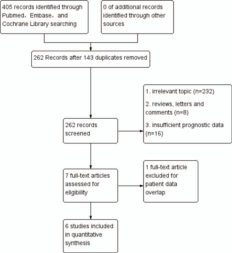
Eligible relevant literatures were retrieved by searching PubMed, the Cochrane Library, Embase through December 2016. Articles that comparing different PKM2 expression levels in human breast cancer tissues and prognostic significance were included. Software RevMan 5.3 and STATA (Review Manager (RevMan): [Computer program]. Version 5.3. Copenhagen: The Nordic Cochrane Centre, The Cochrane Collaboration, 2014.
StataCorp. 2011. Stata Statistical Software: Release 12. College Station, TX: StataCorp LP) were applied to analyze the outcomes. Pooled results were presented in hazardous ratios (HRs) of 5-year overall survival (OS), progression-free survival (PFS), and odds ratios (ORs) of clinicopathological features with 95% confidence intervals.
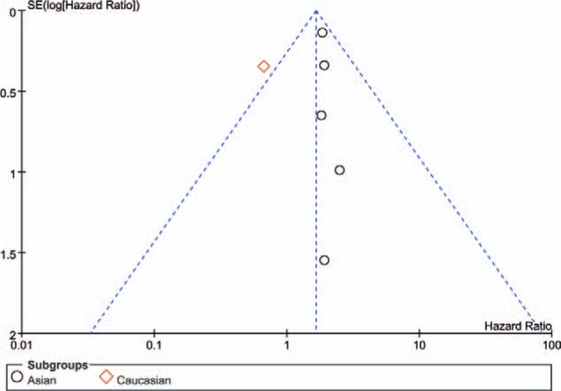
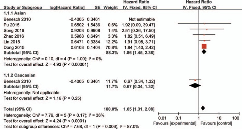
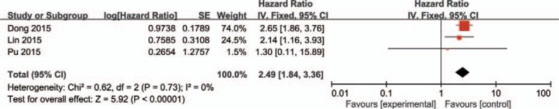
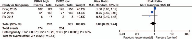
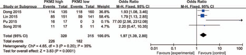
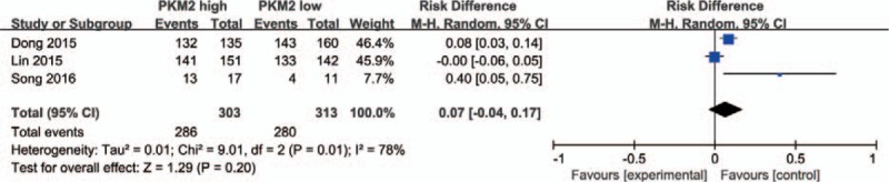
Data from 6 involved studies with 895 patients were summarized. Breast cancer patients with high PKM2 had a worse OS (pooled HR = 1.65, 95% CI = 1.31-2.08, P < .001) and PFS (pooled HR = 2.49, 95% CI = 1.84-3.36, P < .00001). High PKM2 expression is related to lymph node metastasis (N1+N2+N3 vs N0, OR = 1.97, 95%CI = 1.39-2.80, P = .0001). The outcome stability was verified via sensitivity analysis. But elevated PKM2 expression was not correlated to tumor stage (T2+T3 vs T1, pooled OR = 0.80, 95% CI = 0.36-1.77, P = .58) and differential grade (G2+G3 vs G1, OR = 2.74, 95%CI = 0.76-9.84, P = .12). No publication bias was found in the included studies for OS (Begg test, P = .260; Egger test, P = .747).
High PKM2 expression denotes worse OS and PFS in breast cancer patients, and correlate with the lymph node metastasis. However, there is no evidence for the impact of PKM2 expression on T stage and tumor differentiation.
众多研究报道,癌症中丙酮酸激酶M2亚型(PKM2)表达异常,这表明PKM2在肿瘤的发生和发展中起关键作用。然而,其在乳腺癌中的预后价值仍存在争议。因此,我们进行了这项荟萃分析,以评估PKM2在乳腺癌中的预后意义。
通过检索截至2016年12月的PubMed、Cochrane图书馆、Embase来获取符合条件的相关文献。纳入比较人类乳腺癌组织中不同PKM2表达水平及其预后意义的文章。应用RevMan 5.3软件和STATA(Review Manager(RevMan):[计算机程序]。第5.3版。哥本哈根:北欧Cochrane中心,Cochrane协作网,2014年。STATA:StataCorp。2011年。Stata统计软件:第12版。德克萨斯州大学站:StataCorp LP)分析结果。汇总结果以5年总生存期(OS)、无进展生存期(PFS)的风险比(HRs)以及临床病理特征的优势比(ORs)和95%置信区间呈现。
总结了6项涉及895例患者的研究数据。PKM2高表达的乳腺癌患者总生存期更差(汇总HR = 1.65,95%CI = 1.31 - 2.08,P <.001),无进展生存期也更差(汇总HR = 2.49,95%CI = 1.84 - 3.36,P <.00001)。PKM2高表达与淋巴结转移相关(N1 + N2 + N3与N0相比,OR = 1.97,95%CI = 1.39 - 2.80,P =.0001)。通过敏感性分析验证了结果的稳定性。但PKM2表达升高与肿瘤分期(T2 + T3与T1相比,汇总OR = 0.80,95%CI = 0.36 - 1.77,P =.58)和分化程度(G2 + G3与G1相比,OR = 2.74,95%CI = 0.76 - 9.84,P =.12)无关。纳入的总生存期研究中未发现发表偏倚(Begg检验,P =.260;Egger检验,P =.747)。
PKM2高表达表明乳腺癌患者总生存期和无进展生存期较差,且与淋巴结转移相关。然而,没有证据表明PKM2表达对T分期和肿瘤分化有影响。