Department of Basic and Clinical Neuroscience, Institute of Psychiatry, Psychology and Neuroscience, King's College London, London, United Kingdom.
Developmental Imaging and Biophysics Section, Developmental Neurosciences Program, UCL Great Ormond Street Institute of Child Health, London, United Kingdom.
Epilepsia. 2018 Jan;59(1):226-234. doi: 10.1111/epi.13955. Epub 2017 Nov 18.
Patients with genetic generalized epilepsy (GGE) have subtle morphologic abnormalities of the brain revealed with magnetic resonance imaging (MRI), particularly in the thalamus. However, it is unclear whether morphologic abnormalities of the brain in GGE are a consequence of repeated seizures over the duration of the disease, or are a consequence of treatment with antiepileptic drugs (AEDs), or are independent of these factors. Therefore, we measured brain morphometry in a cohort of AED-naive patients with GGE at disease onset. We hypothesize that drug-naive patients at disease onset have gray matter changes compared to age-matched healthy controls.
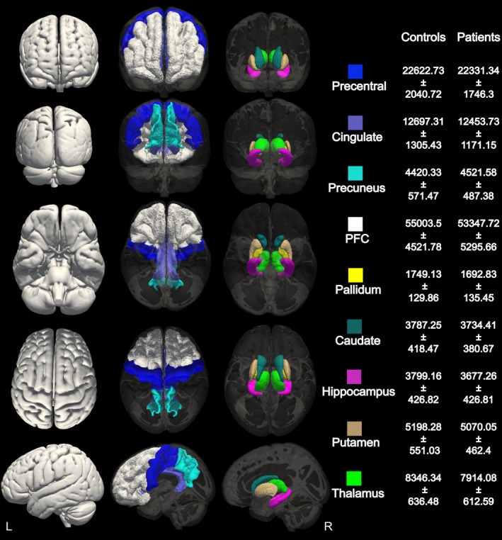
We performed quantitative measures of gray matter volume in the thalamus, putamen, caudate, pallidum, hippocampus, precuneus, prefrontal cortex, precentral cortex, and cingulate in 29 AED-naive patients with new-onset GGE and compared them to 32 age-matched healthy controls. We subsequently compared the shape of any brain structures found to differ in gray matter volume between the groups.
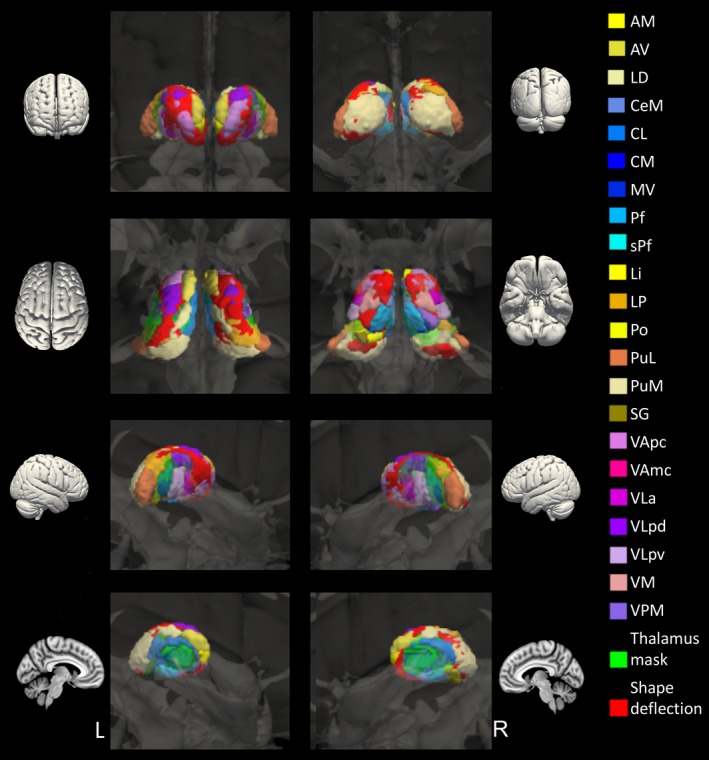
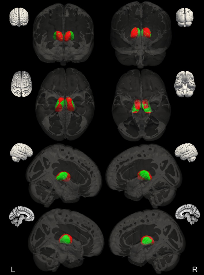
The thalamus was the only structure to show reduced gray matter volume in AED-naive patients with new-onset GGE compared to healthy controls. Shape analysis revealed that the thalamus showed deflation, which was not uniformly distributed, but particularly affected a circumferential strip involving anterior, superior, posterior, and inferior regions with sparing of medial and lateral regions.
Structural abnormalities in the thalamus are present at the initial onset of GGE in AED-naive patients, suggesting that thalamic structural abnormality is an intrinsic feature of GGE and not a consequence of AEDs or disease duration.
患有遗传性全面性癫痫(GGE)的患者通过磁共振成像(MRI)显示出大脑的细微形态异常,尤其是在丘脑。然而,尚不清楚 GGE 患者大脑的形态异常是疾病持续时间内反复癫痫发作的结果,还是抗癫痫药物(AED)治疗的结果,或者是否与这些因素无关。因此,我们在疾病发作时对一组未经 AED 治疗的 GGE 患者进行了脑部形态计量学测量。我们假设,与年龄匹配的健康对照组相比,疾病发作时未经药物治疗的患者有灰质变化。
我们对 29 名新诊断的 GGE 且未接受 AED 治疗的患者的丘脑、壳核、尾状核、苍白球、海马体、楔前叶、前额叶皮质、中央前回和扣带回进行了灰质体积的定量测量,并与 32 名年龄匹配的健康对照组进行了比较。随后,我们比较了两组间灰质体积差异较大的任何脑结构的形状。
与健康对照组相比,新诊断的 GGE 且未接受 AED 治疗的患者的丘脑是唯一灰质体积减少的结构。形态分析显示,丘脑呈萎缩性改变,且分布不均匀,主要影响包括前、上、后和下区域的环形带,而内侧和外侧区域不受影响。
在未经 AED 治疗的新诊断 GGE 患者中,丘脑存在结构异常,这表明丘脑结构异常是 GGE 的固有特征,而不是 AED 或疾病持续时间的结果。