• 文献检索
  • 文档翻译
  • 深度研究
  • 学术资讯
  • Suppr Zotero 插件Zotero 插件
  • 邀请有礼
  • 套餐&价格
  • 历史记录
应用&插件
Suppr Zotero 插件Zotero 插件浏览器插件Mac 客户端Windows 客户端微信小程序
定价
高级版会员购买积分包购买API积分包
服务
文献检索文档翻译深度研究API 文档MCP 服务
关于我们
关于 Suppr公司介绍联系我们用户协议隐私条款
关注我们

Suppr 超能文献

核心技术专利:CN118964589B侵权必究
粤ICP备2023148730 号-1Suppr @ 2026

文献检索

告别复杂PubMed语法,用中文像聊天一样搜索,搜遍4000万医学文献。AI智能推荐,让科研检索更轻松。

立即免费搜索

文件翻译

保留排版,准确专业,支持PDF/Word/PPT等文件格式,支持 12+语言互译。

免费翻译文档

深度研究

AI帮你快速写综述,25分钟生成高质量综述,智能提取关键信息,辅助科研写作。

立即免费体验

电解液pH值对增材制造Ti64合金上电化学沉积羟基磷灰石涂层性能的影响。

Influence of the electrolyte's pH on the properties of electrochemically deposited hydroxyapatite coating on additively manufactured Ti64 alloy.

作者信息

Vladescu Alina, Vranceanu Diana M, Kulesza Slawek, Ivanov Alexey N, Bramowicz Mirosław, Fedonnikov Alexander S, Braic Mariana, Norkin Igor A, Koptyug Andrey, Kurtukova Maria O, Dinu Mihaela, Pana Iulian, Surmeneva Maria A, Surmenev Roman A, Cotrut Cosmin M

机构信息

National Institute for Optoelectronics, Department for Advanced Surface Processing and Analysis by Vacuum Technologies, 409 Atomistilor St., Magurele, RO77125, Romania.

National Research Tomsk Polytechnic University, Lenin Avenue 43, Tomsk, 634050, Russia.

出版信息

Sci Rep. 2017 Dec 1;7(1):16819. doi: 10.1038/s41598-017-16985-z.

DOI:10.1038/s41598-017-16985-z
PMID:29196637
原文链接:https://pmc.ncbi.nlm.nih.gov/articles/PMC5711918/
Abstract

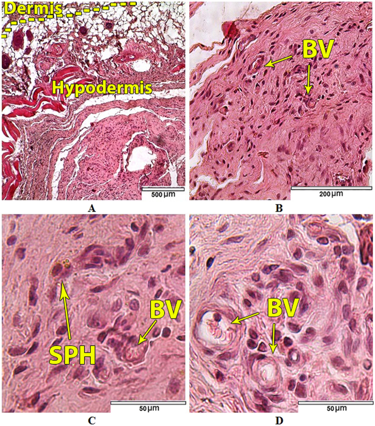
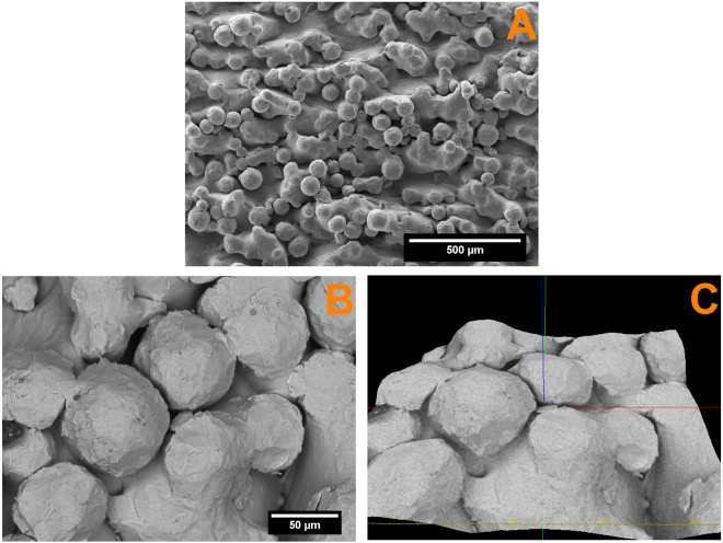
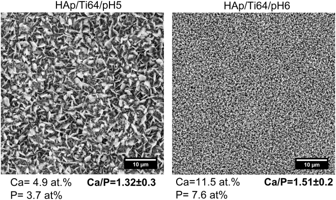
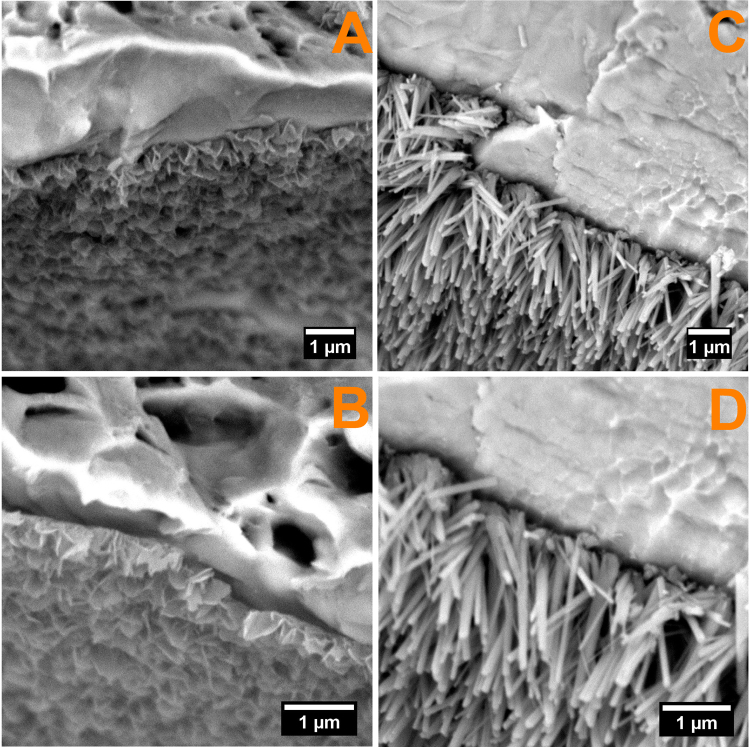
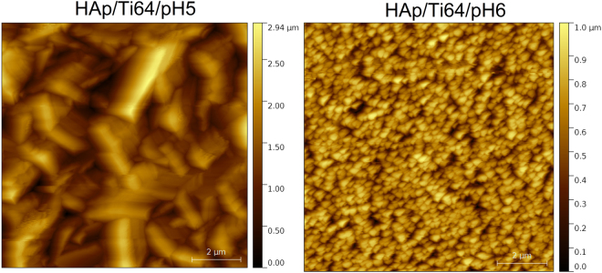
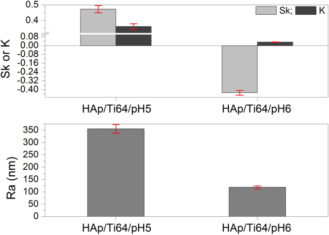
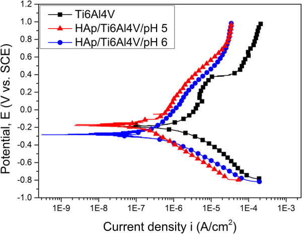
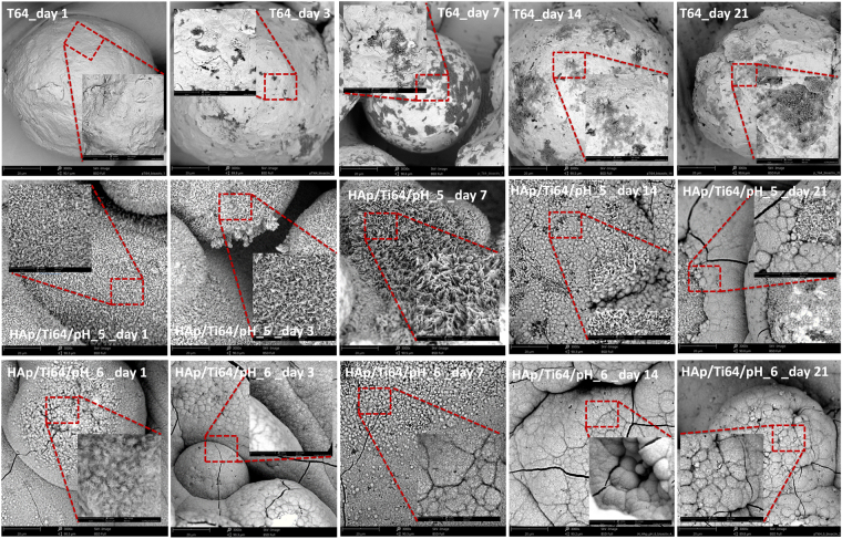
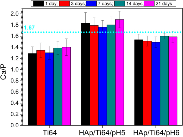
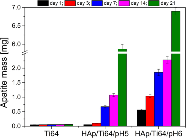
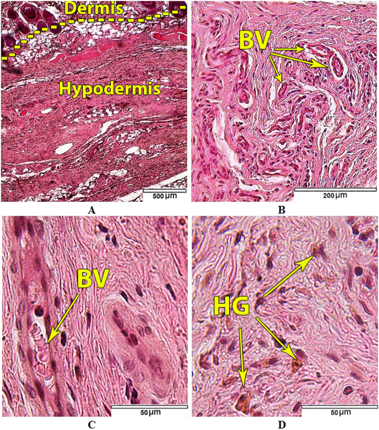
Properties of the hydroxyapatite obtained by electrochemical assisted deposition (ED) are dependent on several factors including deposition temperature, electrolyte pH and concentrations, applied potential. All of these factors directly influence the morphology, stoichiometry, crystallinity, electrochemical behaviour, and particularly the coating thickness. Coating structure together with surface micro- and nano-scale topography significantly influence early stages of the implant bio-integration. The aim of this study is to analyse the effect of pH modification on the morphology, corrosion behaviour and in vitro bioactivity and in vivo biocompatibility of hydroxyapatite prepared by ED on the additively manufactured Ti64 samples. The coatings prepared in the electrolytes with pH = 6 have predominantly needle like morphology with the dimensions in the nanometric scale (30 nm). Samples coated at pH = 6 demonstrated higher protection efficiency against the corrosive attack as compared to the ones coated at pH = 5 (93% against 89%). The in vitro bioactivity results indicated that both coatings have a greater capacity of biomineralization, compared to the uncoated Ti64. Somehow, the coating deposited at pH = 6 exhibited good corrosion behaviour and high biomineralization ability. In vivo subcutaneous implantation of the coated samples into the white rats for up to 21 days with following histological studies showed no serious inflammatory process.

摘要

通过电化学辅助沉积(ED)获得的羟基磷灰石的性能取决于几个因素,包括沉积温度、电解质的pH值和浓度、施加的电势。所有这些因素都直接影响其形态、化学计量、结晶度、电化学行为,尤其是涂层厚度。涂层结构以及表面微观和纳米尺度的形貌显著影响植入物生物整合的早期阶段。本研究的目的是分析pH值改变对通过ED在增材制造的Ti64样品上制备的羟基磷灰石的形态、腐蚀行为、体外生物活性和体内生物相容性的影响。在pH = 6的电解质中制备的涂层主要呈针状形态,尺寸在纳米尺度(约30 nm)。与在pH = 5下涂层的样品相比,在pH = 6下涂层的样品对腐蚀攻击表现出更高的保护效率(约93%对89%)。体外生物活性结果表明,与未涂层的Ti64相比,两种涂层都具有更大的生物矿化能力。不知何故,在pH = 6下沉积的涂层表现出良好的腐蚀行为和高生物矿化能力。将涂层样品皮下植入白色大鼠体内长达21天,随后进行组织学研究,结果显示没有严重的炎症过程。

https://cdn.ncbi.nlm.nih.gov/pmc/blobs/bfbe/5711918/be36329da60b/41598_2017_16985_Fig16_HTML.jpg
https://cdn.ncbi.nlm.nih.gov/pmc/blobs/bfbe/5711918/0c273c2f6826/41598_2017_16985_Fig1_HTML.jpg
https://cdn.ncbi.nlm.nih.gov/pmc/blobs/bfbe/5711918/7b618eddb1ef/41598_2017_16985_Fig2_HTML.jpg
https://cdn.ncbi.nlm.nih.gov/pmc/blobs/bfbe/5711918/8471f6938b83/41598_2017_16985_Fig3_HTML.jpg
https://cdn.ncbi.nlm.nih.gov/pmc/blobs/bfbe/5711918/7e1db96bd256/41598_2017_16985_Fig4_HTML.jpg
https://cdn.ncbi.nlm.nih.gov/pmc/blobs/bfbe/5711918/4f719ce69fdf/41598_2017_16985_Fig5_HTML.jpg
https://cdn.ncbi.nlm.nih.gov/pmc/blobs/bfbe/5711918/f70f90a806b2/41598_2017_16985_Fig6_HTML.jpg
https://cdn.ncbi.nlm.nih.gov/pmc/blobs/bfbe/5711918/cc40b83f5e73/41598_2017_16985_Fig7_HTML.jpg
https://cdn.ncbi.nlm.nih.gov/pmc/blobs/bfbe/5711918/2d8a3d99064a/41598_2017_16985_Fig8_HTML.jpg
https://cdn.ncbi.nlm.nih.gov/pmc/blobs/bfbe/5711918/425bde149e01/41598_2017_16985_Fig9_HTML.jpg
https://cdn.ncbi.nlm.nih.gov/pmc/blobs/bfbe/5711918/1ed15bc23454/41598_2017_16985_Fig10_HTML.jpg
https://cdn.ncbi.nlm.nih.gov/pmc/blobs/bfbe/5711918/bcea88adb8ab/41598_2017_16985_Fig11_HTML.jpg
https://cdn.ncbi.nlm.nih.gov/pmc/blobs/bfbe/5711918/150bb570a7ad/41598_2017_16985_Fig12_HTML.jpg
https://cdn.ncbi.nlm.nih.gov/pmc/blobs/bfbe/5711918/b743460a3405/41598_2017_16985_Fig13_HTML.jpg
https://cdn.ncbi.nlm.nih.gov/pmc/blobs/bfbe/5711918/ac2219d59646/41598_2017_16985_Fig14_HTML.jpg
https://cdn.ncbi.nlm.nih.gov/pmc/blobs/bfbe/5711918/e9fd3e43947e/41598_2017_16985_Fig15_HTML.jpg
https://cdn.ncbi.nlm.nih.gov/pmc/blobs/bfbe/5711918/be36329da60b/41598_2017_16985_Fig16_HTML.jpg

相似文献

1
Influence of the electrolyte's pH on the properties of electrochemically deposited hydroxyapatite coating on additively manufactured Ti64 alloy.电解液pH值对增材制造Ti64合金上电化学沉积羟基磷灰石涂层性能的影响。
Sci Rep. 2017 Dec 1;7(1):16819. doi: 10.1038/s41598-017-16985-z.
2
Preparation and corrosion resistance of magnesium phytic acid/hydroxyapatite composite coatings on biodegradable AZ31 magnesium alloy.可生物降解AZ31镁合金上植酸/羟基磷灰石复合涂层的制备及其耐蚀性
J Mater Sci Mater Med. 2017 Jun;28(6):82. doi: 10.1007/s10856-017-5876-9. Epub 2017 Apr 19.
3
Biocompatibility of hydroxyapatite coatings deposited by pulse electrodeposition technique on the Nitinol superelastic alloy.通过脉冲电沉积技术在镍钛诺超弹性合金上沉积的羟基磷灰石涂层的生物相容性。
Mater Sci Eng C Mater Biol Appl. 2017 Jul 1;76:278-286. doi: 10.1016/j.msec.2017.03.064. Epub 2017 Mar 10.
4
Electrochemically assisted deposition of hydroxyapatite on Ti6Al4V substrates covered by CVD diamond films - Coating characterization and first cell biological results.在化学气相沉积(CVD)金刚石薄膜覆盖的Ti6Al4V基底上进行羟基磷灰石的电化学辅助沉积——涂层表征及首次细胞生物学结果
Mater Sci Eng C Mater Biol Appl. 2016 Feb;59:624-635. doi: 10.1016/j.msec.2015.10.063. Epub 2015 Oct 22.
5
Deposition of nanostructured fluorine-doped hydroxyapatite-polycaprolactone duplex coating to enhance the mechanical properties and corrosion resistance of Mg alloy for biomedical applications.沉积纳米结构的氟掺杂羟基磷灰石-聚己内酯双相涂层以增强镁合金在生物医学应用中的力学性能和耐腐蚀性。
Mater Sci Eng C Mater Biol Appl. 2016 Mar;60:526-537. doi: 10.1016/j.msec.2015.11.057. Epub 2015 Nov 22.
6
A biodegradable AZ91 magnesium alloy coated with a thin nanostructured hydroxyapatite for improving the corrosion resistance.一种涂覆有薄纳米结构羟基磷灰石的可生物降解AZ91镁合金,用于提高耐腐蚀性。
Mater Sci Eng C Mater Biol Appl. 2017 Jun 1;75:95-103. doi: 10.1016/j.msec.2017.02.033. Epub 2017 Feb 9.
7
The synergistic effect of hierarchical micro/nano-topography and bioactive ions for enhanced osseointegration.分层微/纳形貌和生物活性离子协同作用增强骨整合。
Biomaterials. 2013 Apr;34(13):3184-95. doi: 10.1016/j.biomaterials.2013.01.008. Epub 2013 Feb 4.
8
Influence of polyetheretherketone coatings on the Ti-13Nb-13Zr titanium alloy's bio-tribological properties and corrosion resistance.聚醚醚酮涂层对 Ti-13Nb-13Zr 钛合金生物摩擦学性能和耐腐蚀性的影响。
Mater Sci Eng C Mater Biol Appl. 2016 Jun;63:52-61. doi: 10.1016/j.msec.2016.02.043. Epub 2016 Feb 19.
9
Electrophoretic Deposition of Nanocrystalline Calcium Phosphate Coating for Augmenting Bioactivity of Additively Manufactured Ti-6Al-4V.用于增强增材制造Ti-6Al-4V生物活性的纳米晶磷酸钙涂层的电泳沉积
ACS Mater Au. 2021 Nov 22;2(2):132-142. doi: 10.1021/acsmaterialsau.1c00043. eCollection 2022 Mar 9.
10
Corrosion resistance and surface biocompatibility of a microarc oxidation coating on a Mg-Ca alloy.镁钙合金微弧氧化涂层的耐腐蚀和表面生物相容性。
Acta Biomater. 2011 Apr;7(4):1880-9. doi: 10.1016/j.actbio.2010.11.034. Epub 2010 Dec 8.

引用本文的文献

1
Advanced surface modification techniques for titanium implants: a review of osteogenic and antibacterial strategies.钛植入物的先进表面改性技术:成骨和抗菌策略综述
Front Bioeng Biotechnol. 2025 Mar 19;13:1549439. doi: 10.3389/fbioe.2025.1549439. eCollection 2025.
2
Citrus-Fruit-Based Hydroxyapatite Anodization Coatings on Titanium Implants.钛植入物上基于柑橘类水果的羟基磷灰石阳极氧化涂层
Materials (Basel). 2025 Mar 5;18(5):1163. doi: 10.3390/ma18051163.
3
Exploring the Broad Spectrum of Titanium-Niobium Implants and Hydroxyapatite Coatings-A Review.

本文引用的文献

1
Fabrication of Metallic Biomedical Scaffolds with the Space Holder Method: A Review.采用空间保持法制备金属生物医学支架:综述
Materials (Basel). 2014 May 6;7(5):3588-3622. doi: 10.3390/ma7053588.
2
Expression of Concern: Nanodimensional and Nanocrystalline Apatites and Other Calcium Orthophosphates in Biomedical Engineering, Biology and Medicine. Materials 2009, 2, 1975-2045.关注声明:生物医学工程、生物学与医学中的纳米尺寸和纳米晶磷灰石及其他正磷酸钙。《材料》2009年,第2卷,第1975 - 2045页。
Materials (Basel). 2016 Sep 2;9(9):752. doi: 10.3390/ma9090752.
3
Additively Manufactured Scaffolds for Bone Tissue Engineering and the Prediction of their Mechanical Behavior: A Review.
探索钛铌植入物和羟基磷灰石涂层的广泛领域——综述
Materials (Basel). 2024 Dec 19;17(24):6206. doi: 10.3390/ma17246206.
4
Selection Route of Precursor Materials in 3D Printing Composite Filament Development for Biomedical Applications.生物医学应用3D打印复合长丝开发中前驱体材料的选择途径
Materials (Basel). 2023 Mar 15;16(6):2359. doi: 10.3390/ma16062359.
5
Modulated Laser Cladding of Implant-Type Coatings by Bovine-Bone-Derived Hydroxyapatite Powder Injection on Ti6Al4V Substrates-Part I: Fabrication and Physico-Chemical Characterization.通过在Ti6Al4V基体上注射牛骨衍生的羟基磷灰石粉末对植入型涂层进行调制激光熔覆 - 第一部分:制备与物理化学表征
Materials (Basel). 2022 Nov 11;15(22):7971. doi: 10.3390/ma15227971.
6
Evaluation of the Cathodic Electrodeposition Effectiveness of the Hydroxyapatite Layer Used in Surface Modification of Ti6Al4V-Based Biomaterials.用于Ti6Al4V基生物材料表面改性的羟基磷灰石层的阴极电沉积有效性评估。
Materials (Basel). 2022 Oct 6;15(19):6925. doi: 10.3390/ma15196925.
7
Electro-deposition of bactericidal and corrosion-resistant hydroxyapatite nanoslabs.杀菌且耐腐蚀的羟基磷灰石纳米片的电沉积
RSC Adv. 2019 Apr 9;9(20):11170-11178. doi: 10.1039/c9ra00811j.
8
Effect of Deep Cryogenic Treatment on Corrosion Behavior of AISI H13 Die Steel.深冷处理对AISI H13模具钢腐蚀行为的影响
Materials (Basel). 2021 Dec 18;14(24):7863. doi: 10.3390/ma14247863.
9
Nanohydroxyapatite Electrodeposition onto Electrospun Nanofibers: Technique Overview and Tissue Engineering Applications.纳米羟基磷灰石在电纺纳米纤维上的电沉积:技术概述与组织工程应用
Bioengineering (Basel). 2021 Oct 22;8(11):151. doi: 10.3390/bioengineering8110151.
10
Physical Properties, Spectroscopic, Microscopic, X-ray, and Chemometric Analysis of Starch Films Enriched with Selected Functional Additives.富含选定功能添加剂的淀粉膜的物理性质、光谱、显微镜、X射线和化学计量学分析
Materials (Basel). 2021 May 20;14(10):2673. doi: 10.3390/ma14102673.
用于骨组织工程的增材制造支架及其力学行为预测:综述
Materials (Basel). 2017 Jan 10;10(1):50. doi: 10.3390/ma10010050.
4
Metallic powder-bed based 3D printing of cellular scaffolds for orthopaedic implants: A state-of-the-art review on manufacturing, topological design, mechanical properties and biocompatibility.用于骨科植入物的基于金属粉末床的细胞支架3D打印:关于制造、拓扑设计、力学性能和生物相容性的最新综述
Mater Sci Eng C Mater Biol Appl. 2017 Jul 1;76:1328-1343. doi: 10.1016/j.msec.2017.02.094. Epub 2017 Feb 24.
5
The Influence of Electrolytic Concentration on the Electrochemical Deposition of Calcium Phosphate Coating on a Direct Laser Metal Forming Surface.电解液浓度对直接激光金属成型表面磷酸钙涂层电化学沉积的影响。
Int J Anal Chem. 2017;2017:8610858. doi: 10.1155/2017/8610858. Epub 2017 Jan 29.
6
In vivo vascularization of MSC-loaded porous hydroxyapatite constructs coated with VEGF-functionalized collagen/heparin multilayers.负载间充质干细胞的多孔羟基磷灰石构建体在体内的血管化,构建体表面涂覆有血管内皮生长因子功能化的胶原蛋白/肝素多层膜。
Sci Rep. 2016 Jan 22;6:19871. doi: 10.1038/srep19871.
7
A review of hydroxyapatite-based coating techniques: Sol-gel and electrochemical depositions on biocompatible metals.基于羟基磷灰石的涂层技术综述:生物相容性金属上的溶胶-凝胶和电化学沉积法
J Mech Behav Biomed Mater. 2016 Apr;57:95-108. doi: 10.1016/j.jmbbm.2015.11.031. Epub 2015 Dec 6.
8
Calcium orthophosphate deposits: Preparation, properties and biomedical applications.正磷酸钙沉积物:制备、性质及生物医学应用。
Mater Sci Eng C Mater Biol Appl. 2015 Oct;55:272-326. doi: 10.1016/j.msec.2015.05.033. Epub 2015 May 13.
9
3D Printing of Scaffolds for Tissue Regeneration Applications.用于组织再生应用的支架的3D打印
Adv Healthc Mater. 2015 Aug 26;4(12):1742-62. doi: 10.1002/adhm.201500168. Epub 2015 Jun 10.
10
3D printing of porous hydroxyapatite scaffolds intended for use in bone tissue engineering applications.用于骨组织工程应用的多孔羟基磷灰石支架的 3D 打印。
Mater Sci Eng C Mater Biol Appl. 2015 Feb;47:237-47. doi: 10.1016/j.msec.2014.11.024. Epub 2014 Nov 8.