Hu Shao-Hua, Feng Hong, Xu Ting-Ting, Zhang Hao-Rong, Zhao Zhi-Yong, Lai Jian-Bo, Xu Dong-Rong, Xu Yi
Department of Psychiatry, First Affiliated Hospital, Zhejiang University School of Medicine, Hangzhou.
The Key Laboratory of Mental Disorder's Management of Zhejiang Province, Hangzhou.
Neuropsychiatr Dis Treat. 2017 Nov 21;13:2829-2836. doi: 10.2147/NDT.S144972. eCollection 2017.
Structural studies have reported anorexia nervosa (AN) patients with abnormal gray matter in several brain regions and dysfunction in some connected neural circuits. However, the role of white matter (WM) in AN patients has rarely been investigated. The present study aimed to assess alterations in WM microstructure of the entire brain in females with AN using a voxel-based method on diffusion tensor imaging (DTI) data.
The study enrolled 8 female patients with AN and 14 age-matched females as controls (CW). The DTI data was collected from each subject to calculate the fractional anisotropy (FA) maps of the whole brain by the DTI-Studio software. Subsequently, a 2-sample -test (<0.05, corrected) was performed to detect the difference in FA maps of AN and CW group, and a Pearson's correlation analyzed the relationship between mean FA value of brain regions and body mass index (BMI).
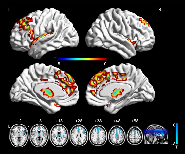
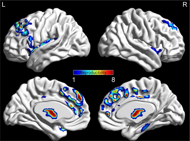
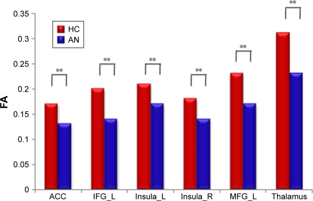
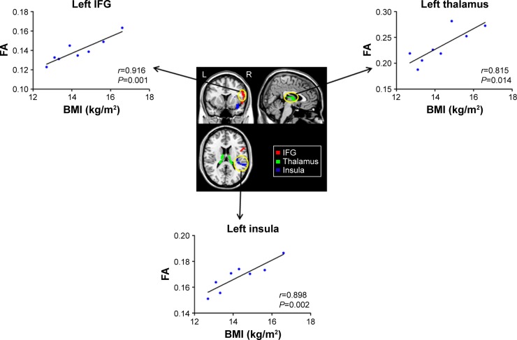
Compared with CW, AN patients revealed a significant decrease in FA maps in the left superior frontal gyrus, medial frontal gyrus, anterior cingulate cortex, middle frontal gyrus, inferior frontal gyrus, thalamus, and bilateral insula. Moreover, significantly positive correlations were established between the mean FA value of the left inferior frontal gyrus, insula as well as thalamus and BMI in AN patients.
Our findings supported the presence of WM abnormality in patients with AN. The significant differences of FA maps, in patients with AN, were associated with their aberrant BMI. The results further improved our understanding of the pathophysiological mechanisms underlying AN.
结构研究报告称,神经性厌食症(AN)患者在几个脑区存在灰质异常,且一些相连神经回路功能失调。然而,白质(WM)在AN患者中的作用鲜有研究。本研究旨在使用基于体素的方法对扩散张量成像(DTI)数据进行分析,评估AN女性患者全脑白质微结构的改变。
本研究纳入8例AN女性患者和14例年龄匹配的女性作为对照(CW)。收集每位受试者的DTI数据,通过DTI-Studio软件计算全脑的分数各向异性(FA)图。随后,进行双样本t检验(<0.05,校正)以检测AN组和CW组FA图的差异,并采用Pearson相关性分析脑区平均FA值与体重指数(BMI)之间的关系。
与CW相比,AN患者在左侧额上回、额中回、前扣带回皮质、额下回、丘脑及双侧岛叶的FA图显著降低。此外,AN患者左侧额下回、岛叶及丘脑的平均FA值与BMI之间存在显著正相关。
我们的研究结果支持AN患者存在白质异常。AN患者FA图的显著差异与他们异常的BMI相关。这些结果进一步加深了我们对AN潜在病理生理机制的理解。