Department of Medicine, University of Crete, Heraklion, Greece.
Computational Bio-Medicine Laboratory, Institute of Computer Science, Foundation for Research and Technology-Hellas, Heraklion, Greece.
Biomed Res Int. 2017;2017:8569328. doi: 10.1155/2017/8569328. Epub 2017 Oct 31.
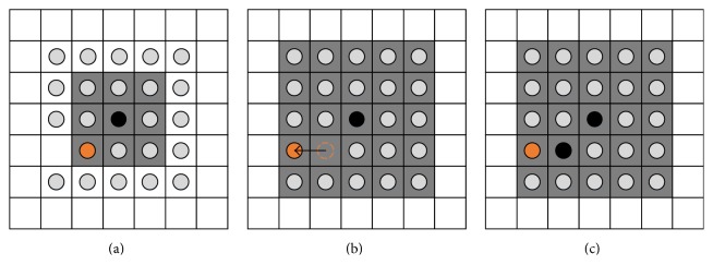
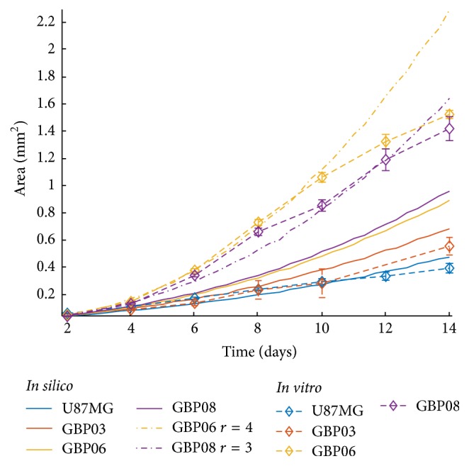
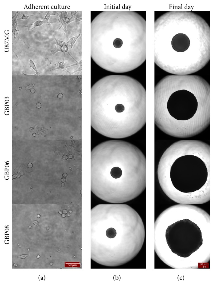
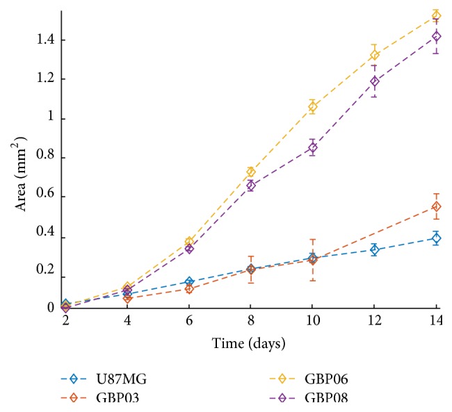
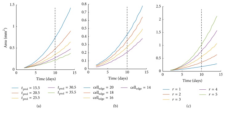
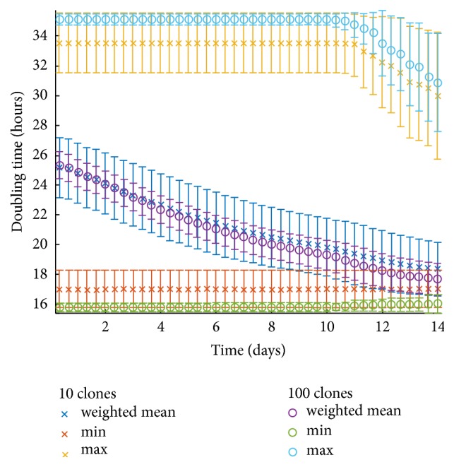
The application of accurate cancer predictive algorithms validated with experimental data is a field concerning both basic researchers and clinicians, especially regarding a highly aggressive form of cancer, such as Glioblastoma. In an aim to enhance prediction accuracy in realistic patient-specific environments, accounting for both inter- and intratumoral heterogeneity, we use patient-derived Glioblastoma cells from different patients. We focus on cell proliferation using experiments to estimate cell doubling times and sizes for established primary Glioblastoma cell lines. A preclinically driven mathematical model parametrization is accomplished by taking into account the experimental measurements. As a control cell line we use the well-studied U87MG cells. Both and results presented support that the variance between tumor staging can be attributed to the differential proliferative capacity of the different Glioblastoma cells. More specifically, the together with the overall proliferation reflected in both the and the can predict the evolution of different Glioblastoma cell lines growing under the same conditions. Undoubtedly, additional imaging techniques capable of providing spatial information of tumor cell physiology and microenvironment will enhance our understanding regarding Glioblastoma nature and verify and further improve our predictability.
应用经过实验数据验证的精确癌症预测算法是一个涉及基础研究人员和临床医生的领域,特别是对于高度侵袭性的癌症,如神经胶质瘤。为了提高在现实患者特定环境中的预测准确性,考虑到肿瘤内和肿瘤间的异质性,我们使用来自不同患者的患者来源的神经胶质瘤细胞。我们专注于细胞增殖,使用实验来估计已建立的原发性神经胶质瘤细胞系的细胞倍增时间和大小。通过考虑实验测量值,完成了基于临床前的数学模型参数化。作为对照细胞系,我们使用了研究充分的 U87MG 细胞。和结果都支持这样一种观点,即肿瘤分期之间的差异可以归因于不同神经胶质瘤细胞的不同增殖能力。更具体地说,和整体增殖,反映在和中,可以预测在相同条件下生长的不同神经胶质瘤细胞系的进化。毫无疑问,能够提供肿瘤细胞生理学和微环境空间信息的额外成像技术将增强我们对神经胶质瘤性质的理解,并验证和进一步提高我们的预测能力。