Research Institute of Computer Vision and Robotics, University of Girona, Spain; Computer Science Department, Faculty of Computers and Information, Assiut University, Egypt.
Research Institute of Computer Vision and Robotics, University of Girona, Spain.
Neuroimage Clin. 2017 Nov 20;17:607-615. doi: 10.1016/j.nicl.2017.11.015. eCollection 2018.
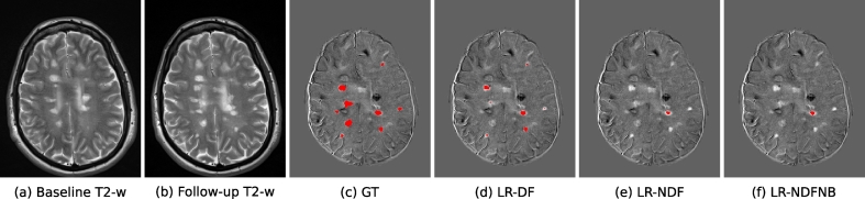
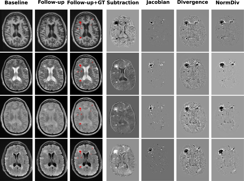
Longitudinal magnetic resonance imaging (MRI) analysis has an important role in multiple sclerosis diagnosis and follow-up. The presence of new T2-w lesions on brain MRI scans is considered a prognostic and predictive biomarker for the disease. In this study, we propose a supervised approach for detecting new T2-w lesions using features from image intensities, subtraction values, and deformation fields (DF).
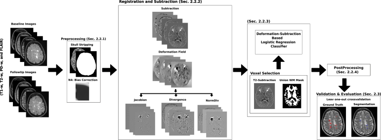
One year apart multi-channel brain MRI scans were obtained for 60 patients, 36 of them with new T2-w lesions. Images from both temporal points were preprocessed and co-registered. Afterwards, they were registered using multi-resolution affine registration, allowing their subtraction. In particular, the DFs between both images were computed with the Demons non-rigid registration algorithm. Afterwards, a logistic regression model was trained with features from image intensities, subtraction values, and DF operators. We evaluated the performance of the model following a leave-one-out cross-validation scheme.
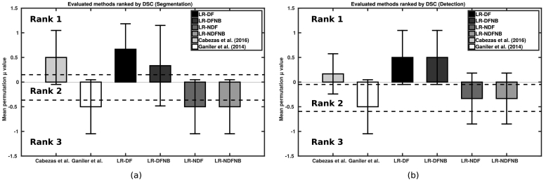
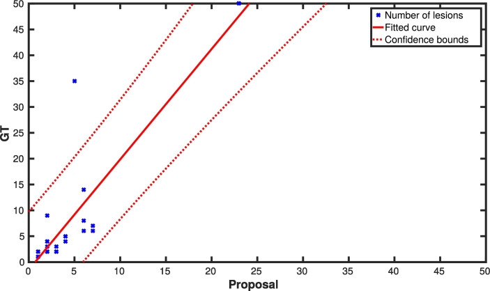
In terms of detection, we obtained a mean Dice similarity coefficient of 0.77 with a true-positive rate of 74.30% and a false-positive detection rate of 11.86%. In terms of segmentation, we obtained a mean Dice similarity coefficient of 0.56. The performance of our model was significantly higher than state-of-the-art methods.
The performance of the proposed method shows the benefits of using DF operators as features to train a supervised learning model. Compared to other methods, the proposed model decreases the number of false-positives while increasing the number of true-positives, which is relevant for clinical settings.
纵向磁共振成像(MRI)分析在多发性硬化症的诊断和随访中具有重要作用。脑 MRI 扫描上新出现的 T2 加权病变被认为是该疾病的预后和预测生物标志物。在这项研究中,我们提出了一种使用来自图像强度、差值和变形场(DF)的特征来检测新 T2 加权病变的监督方法。
对 60 名患者的一年期多通道脑 MRI 扫描进行了研究,其中 36 名患者出现了新的 T2 加权病变。来自两个时间点的图像进行了预处理和配准。然后,使用多分辨率仿射配准对其进行配准,允许它们相减。特别是,使用 Demons 非刚性配准算法计算了两幅图像之间的 DF。然后,使用来自图像强度、差值和 DF 算子的特征训练逻辑回归模型。我们采用留一交叉验证方案评估模型的性能。
在检测方面,我们获得了 0.77 的平均骰子相似系数,真阳性率为 74.30%,假阳性检测率为 11.86%。在分割方面,我们获得了 0.56 的平均骰子相似系数。我们的模型性能明显优于最新方法。
所提出方法的性能表明了使用 DF 算子作为特征来训练监督学习模型的优势。与其他方法相比,该模型减少了假阳性的数量,同时增加了真阳性的数量,这对于临床环境很重要。